Diagnosis and Subclassification of Acute Lymphoblastic Leukemia

Sabina Chiaretti1, Gina Zini2 and Renato Bassan3

1 Division of Hematology, Department of Cellular Biotechnologies and Hematology, “Sapienza” University of Rome, Rome, Italy
2 Hematology, Catholic University Sacred Heart Policlinico Gemelli, Rome, Italy
3 Hematology and Bone Marrow Transplant Unit, Ospedale dell’Angelo e SS. Giovanni e Paolo, Mestre-Venezia, Italy

Corresponding author: Sabina Chiaretti, Division of Hematology, Department of Cellular Biotechnologies and Hematology,“Sapienza” University of Rome, Via Benevento 6, 00161, Rome, Italy. E-mail: chiaretti@bce.uniroma1.it 

Published: October 24, 2014
Received: September 18, 2014
Accepted: October 20, 2014
Meditter J Hematol Infect Dis 2014, 6(1): e2014073, DOI 10.4084/MJHID.2014.073
This article is available on PDF format at:

This is an Open Access article distributed under the terms of the Creative Commons Attribution License
(http://creativecommons.org/licenses/by/2.0), which permits unrestricted use, distribution, and reproduction in any medium, provided the original work is properly cited.


Abstract

Acute lymphoblastic leukemia (ALL) is a disseminated malignancy of B- or T-lymphoblasts which imposes a rapid and accurate diagnostic process to support an optimal risk-oriented therapy and thus increase the curability rate. The need for a precise diagnostic algorithm is underlined by the awareness that both ALL therapy and related success rates may vary greatly between ALL subsets, from standard chemotherapy in patients with standard-risk ALL, to allotransplantation (SCT) and targeted therapy in high-risk patients and cases expressing suitable biological targets, respectively. This review summarizes how best to identify ALL and the most relevant ALL subsets. 

Introduction

Current standards for acute lymphoblastic leukemia (ALL) diagnosis integrate the study of cell morphology, immunophenotype and genetics/cytogenetics as detailed in the 2008 WHO classification of lymphoid neoplasms.[1] The classification originally suggested by the FAB group is no longer followed.[2,3] The FAB classification was clinically useful since it permitted recognition of probable Burkitt lymphoma in leukemic phase, but it has now been replaced by the WHO classification. Lymphoid neoplasms are assigned, in the most recent WHO classification, to two principal categories: neoplasms derived from B- and T-lineage lymphoid precursors and those derived from mature B, T or NK cells. ALL belongs to the first of these major groups, designated B- or T-lymphoblastic leukemia/lymphoma[4] and including three principal categories: B-lymphoblastic leukemia/lymphoma not otherwise specified, B-lymphoblastic leukemia/lymphoma with recurrent cytogenetic alterations and T-lymphoblastic leukemia/lymphoma. The designation of leukemia/lymphoma reflects the principle that these neoplasms should be classified on the basis of their biological and molecular characteristics, regardless of the sites of involvement. The leukemic variant shows diffuse involvement of the peripheral blood and the bone marrow, while lymphoma is confined to nodal or extranodal sites, with no or minimal involvement of the bone marrow. In the leukemic form, by definition, the bone marrow must contain at least 20% blast cells. A purely leukemic presentation is most typical of B-lineage ALL (85%), while cases of T-lineage disease often present with an associated lymphomatous mass in the mediastinum or other sites.

Diagnostic Morphology and Cytochemistry

A morphological bone marrow assessment represents the first step in the diagnostic pathway, for the primary diagnosis of ALL and for the differentiation from acute myeloid leukemia (AML),[5] since ALL, by definition, always presents with bone marrow involvement. Table 1[6] shows the morphological criteria that are useful for distinguishing between myeloblasts and lymphoblasts, however remembering the limits of morphology in ALL, for which flow cytometry analysis represents the diagnostic gold standard for both the identification of cell lineage and the definition of subset. The morphology of leukemic cells in the peripheral blood can be significantly different from that of the bone marrow, which is always indispensable.

Table 1 
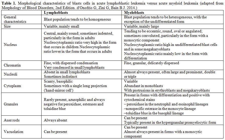
Table 1. Morphological characteristics of blasts cells in acute lymphoblastic leukemia versus acute myeloid leukemia (adapted from Morphology of Blood Disorders, 2nd Edition. d'Onofrio G, Zini G, Bain B.J. 2014).

From the morphological point of view, there are no reproducible criteria to distinguish between B- and T-lineage ALL. It can also be difficult to distinguish B-lineage lymphoblasts from normal B-lineage lymphoid precursors, known as hematogones, which are observed in the peripheral blood in various conditions, including primary myelofibrosis and in children in the phase of recovery following chemotherapy.[7] Hematogones typically have an even higher nucleocytoplasmic ratio than lymphoblasts, with more homogeneous chromatin and a complete absence of visible nucleoli. Hematogones can also express the CD10 antigen, but can be distinguished from blast cells of B ALL by other immunophenotypic features, being characterised by regular, orderly acquisition and loss of B-lineage antigens; they can also be distinguished from mature lymphocytes by their weak expression of CD45 and, sometimes, by the expression of CD34.[7]
The bone marrow morphology of ALL is however quite variable as previously indicated in the FAB classification (Figures 1-2). Rare morphological variants are: ALL with “hand-mirror cells”, i.e. the shape of the cells resembles a hand mirror or a tennis racquet (Figure 2A); granular ALL, with presence of azurophilic cytoplasmic granules which vary in number, size and shape. Cytochemically, these blasts have negative peroxidase reactions and variable periodic acid-Schiff (PAS) positivity; Sudan black B is sometimes weakly positive;[8] ALL with mature cells that are nearly indistinguishable from mature lymphoid neoplasms and require expert observers for accurate morphological identification;[9] ALL associated with hypereosinophilia (Figure 2B). By definition, ALL blasts are negative for myeloperoxidase (MPO) (Figure 2C) and other myeloid cytochemical reactions. According to the FAB criteria, acute (leukemias with at least 3% MPO-positive blasts in BM should be classified as myeloid. However, low level MPO positivity without expression of other myeloid markers is detectable by means of electron microscopy in rare ALL cases. True MPO+ ALL is discussed below in the mixed lineage acute leukemias section. The acid phosphatase reaction correlates with the lysosome content; it is useful for identifying T-ALL blasts which show focal paranuclear positivity in more than 80% of cases. Lymphoblasts may react with non-specific esterases with a strong positivity in the Golgi zone with variable inhibition with sodium fluoride. The B lymphoblasts in FAB L3/Burkitt ALL show an intense cytoplasmic positivity to methyl green pyronine, while the vacuoles stain strongly with Oil red O, thus demonstrating their lipid content. The role of cytochemistry in differentiating ALL from AML is limited and is mainly of historical interest, since these tests have now been superseded by the far more objective results provided by the immunophenotyping.

Figure 1 
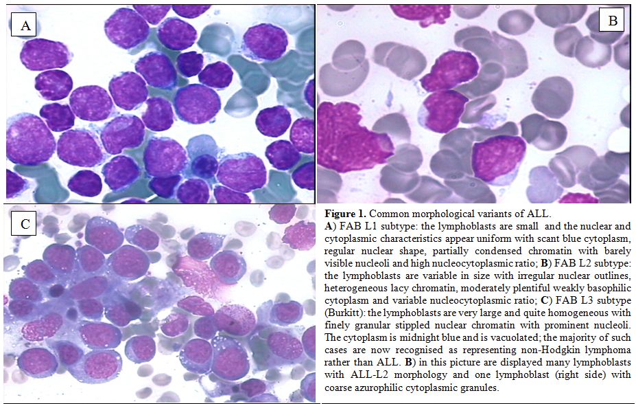
Figure 1. Common morphological variants of ALL.
A) FAB L1 subtype: the lymphoblasts are small  and the nuclear and cytoplasmic characteristics appear uniform with scant blue cytoplasm, regular nuclear shape, partially condensed chromatin with barely visible nucleoli and high nucleocytoplasmic ratio; B) FAB L2 subtype: the lymphoblasts are variable in size with irregular nuclear outlines, heterogeneous lacy chromatin, moderately plentiful weakly basophilic cytoplasm and variable nucleocytoplasmic ratio; C) FAB L3 subtype (Burkitt): the lymphoblasts are very large and quite homogeneous with finely granular stippled nuclear chromatin with prominent nucleoli. The cytoplasm is midnight blue and is vacuolated; the majority of such cases are now recognised as representing non-Hodgkin lymphoma rather than ALL. B) in this picture are displayed many lymphoblasts with ALL-L2 morphology and one lymphoblast (right side) with coarse azurophilic cytoplasmic granules.

Figure 2 
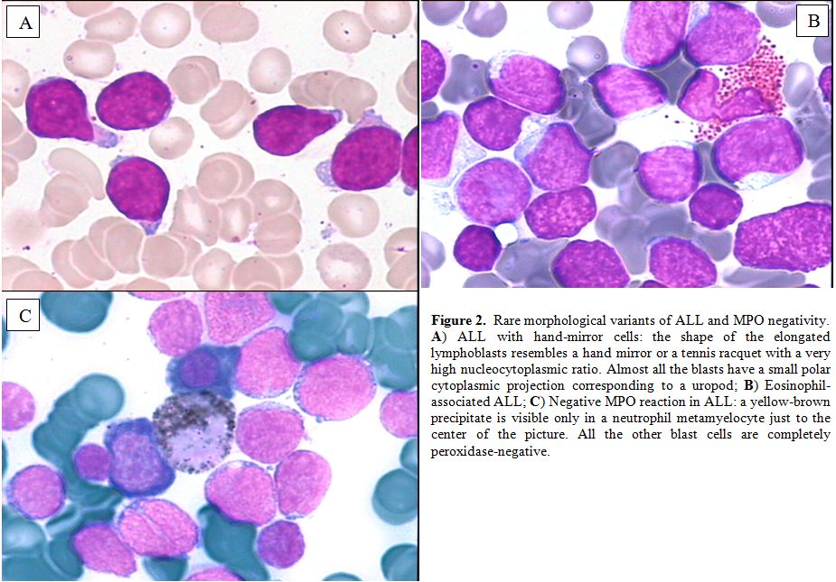
Figure 2. Rare morphological variants of ALL and MPO negativity.
A) ALL with hand-mirror cells: the shape of the elongated lymphoblasts resembles a hand mirror or a tennis racquet with a very high nucleocytoplasmic ratio. Almost all the blasts have a small polar cytoplasmic projection corresponding to a uropod;
B) Eosinophil-associated ALL; C) Negative MPO reaction in ALL: a yellow-brown precipitate is visible only in a neutrophil metamyelocyte just to the center of the picture. All the other blast cells are completely  peroxidase-negative.


Diagnostic Morphology and Cytochemistry

Immunophenotyping by means of multi-channel flow cytometry (MFC) has become the standard procedure for ALL diagnosis and subclassification, and was also developed as useful tool for the detection and monitoring of minimal residual disease (MRD, reviewed elsewhere in this issue). The consensus by European Group for the Immunological characterization of leukaemias (EGIL) is that a threshold of 20% should be used to define a positive reaction of blast cells to a given monoclonal antibody, except for MPO, CD3, CD79a and TdT, which are considered positive at the 10% level of expression.[10,11] More recently, novel MFC strategies were developed by the EuroFlow consortium to ensure accurate methodologies through all MFC steps, in order to guarantee the reproducibility of diagnostic tests.[12,13] To summarize the diagnostic issue, roughly 75-80% of cases of adult ALL are of B-cell lineage and 20-25% belong to the T-cell lineage.

Immunophenotype of B-lineage ALL
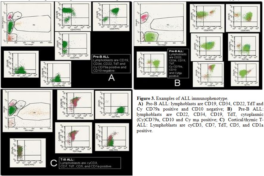
In B-lineage ALL the most important markers for diagnosis, differential diagnosis and subclassification are CD19, CD20, CD22, CD24, and CD79a. The earliest B-lineage markers are CD19, CD22 (membrane and cytoplasm) and CD79a.[14,15] A positive reaction for any two of these three markers, without further differentiation markers, identifies pro-B ALL (EGIL B-I subtype) (Figure 3A). The presence of CD10 antigen (CALLA) defines the "common" ALL subgroup (EGIL B-II subtype). Cases with additional identification of cytoplasmic heavy mu chain constitute the pre-B group (EGIL B-III subtype) (Figure 3B), whereas the presence of surface immunoglobulin light chains defines mature B-ALL (EGIL B-IV subtype).
Among other B-cell markers, B-I and B-II ALL are often CD24 positive and 4G7 (pro- and pre-B surrogate light chain specific MoAb) positive;[16] surface CD20 and CD22 are variably positive beyond stage B-I; CD13 and CD33 myeloid/cross lineage antigen can be expressed, as well as the CD34 stem cell antigen, particularly in Ph+ (Philadelphia chromosome-positive) ALL (often B-II with CD34, CD38, CD25 and CD13/33), but myeloid-specific CD117 should not be present and can be used to differentiate further between ALL and rare myeloid leukaemias with negative MPO expression. Pro-B ALL with t(4;11)/MLL rearrangements is most often myeloid antigen-positive disease (including expression of CD15). TdT expression is usually lost in B-IV subgroup. T-cell markers are usually not expressed in B-lineage ALL but a CD19+ subset is concurrently CD2+. Loss of surface adhesion molecules has been described.[17]
Figure 3
Figure 3. Examples of ALL immunophenotype.
A)  Pro-B ALL: lymphoblasts are CD19, CD34, CD22, TdT and Cy CD79a positive and CD10 negative;
B)  Pre-B ALL: lymphoblasts are CD22, CD34, CD19, TdT, cytoplasmic (Cy)CD79a, CD10 and Cy mμ positive; C)  Cortical/thymic T-ALL: Lymphoblasts are cyCD3, CD7, TdT, CD5, and CD1a positive.

Immunophenotype of T-lineage ALL

T-cell ALL constitutes approximately 25% of all adult cases of ALL. T-cell markers are CD1a, CD2, CD3 (membrane and cytoplasm), CD4, CD5, CD7 and CD8. CD2, CD5 and CD7 antigens are markers of the most immature T-cell cells, but none of them is absolutely lineage-specific, so that the unequivocal diagnosis of T-ALL rests on the demonstration of surface/cytoplasmic CD3. In T-ALL the expression of CD10 is quite common (25%) and not specific; CD34 and myeloid antigens CD13 and/or CD33 can be expressed too. Recognized T-ALL subsets are the following: pro-T EGIL T-I (cCD3+, CD7+), pre-T EGIL T-II (cCD3+, CD7+ and CD5/CD2+), cortical T EGIL T-III (cCD3+, Cd1a+, sCD3+/-) and mature-T EGIL T-IV (cCD3+, sCD3+, CD1a-). Finally, a novel subgroup that was recently characterized is represented by the so called ETP-ALL (Early-T Precursor), which shows characteristic immunophenotypic features, namely lack of CD1a and CD8 expression, weak CD5 expression, and expression of at least one myeloid and/or stem cell marker.[18]

Mixed Phenotype Acute Leukemia

With currently refined diagnostic techniques the occurrence of acute leukemia of ambiguous cell lineage, i.e. mixed phenotype acute leukemia (MPAL) is relatively rare (<4%).[19] These cases express one of the following feature: 1) coexistence of two separate blast cell populations (i.e. T- or B-cell ALL plus either myeloid or monocytic blast cells, 2) single leukemic population of blast cells co-expressing B- or T-cell antigens and myeloid antigens, 3) same plus expression of monocytic antigens. For myelo-monocytic lineage useful diagnostic antigens are MPO or nonspecific esterase, CD11c, CD14, CD64 and lysozyme; for B-lineage CD19 plus CD79a, cytoplasmic CD22 and CD10 (one or two of the latter according to staining intensity of CD19) and for T-lineage cytoplasmic or surface CD3. Recognized entities include Ph+ MPAL (B/myeloid or rarely T/myeloid), t(v;11q23;MLL rearranged MPAL, and genetically uncharacterized B or T/myeloid MPAL. Very rare cases express trilineage involvement (B/T/myeloid). Lack of lineage-specific antigens (MPO, cCD3, cCD22) is observed in the ultra-rare acute undifferentiated leukemia. In a recent review of 100 such cases,[20] 59% were B/myeloid, 35% T/myeloid, 4% B/T lymphoid and 2% B/T/myeloid. Outcome was overall better following ALL rather than AML therapy.

NK Cell ALL

CD56, a marker of natural killer (NK) cell differentiation, defines a rare subgroup of about 3% of adult ALL cases which often display other early T-cell antigens, CD7 CD2 CD5, and sometimes cCD3.[19] True NK ALL is very rare (TdT+, CD56+, other T markers negative, unrearranged TCR genes).[21] This diagnosis rely on the demonstration of early NK-specific CD94 or CD161 antigens.

Differential Diagnosis

With few exceptions, ALL is readily identified by morphological marrow assessment and MFC evaluation, with no need for additional tests, since genetics/cytogenetics and genomics are available at a later stage and cannot be employed for purely diagnostic purposes, even if they add very useful clinical-prognostic information. Differentiation between ALL and AML is initially obtained by excluding reactivity to SBB or MPO stains in ALL cells (<3% positive). On cytochemical evaluation, some rare ALL cases are SBB positive but MPO and chloroacetate esterase are negative. True ALL cases that are immunoreactive to MPO or express detectable levels of MPO mRNA have been described. This can occur in Ph+ ALL and occasionally in T-lineage ALL.[22] Evaluation of CD117 antigen expression should also be carried out.[23] Most ALL cases express the nuclear enzyme Terminal deoxynucleotidyl Transferase (TdT). TdT-negative ALL is uncommonly reported, more in T-ALL, while it is a rule in L3/Burkitt leukemia. Therefore all TdT-negative B-precursor ALL cases must be thoroughly investigated to exclude other aggressive lymphoid neoplasms with leukemic presentation (blastic mantle cell lymphoma, atypical plasmablastic myeloma, other high-grade lymphomas).[24,25]

Diagnostic Cytogenetics

Cytogenetics represents an important step in ALL classification. Conventional karyotyping can be helpful in the identification of recurrent translocations, as well as gain and loss of gross chromosomal material; however, the major limitation of this technique is that in some cases leukemic cells fail to enter metaphase. However, fluorescence in situ hybridization (FISH) can enable the detection and direct visualization of virtually all investigated chromosomal abnormalities in ALL, with a sensitivity of around 99%. Finally, array-comparative genomic hybridization (array-CGH, a-CGH) and single nucleotide polymorphisms (SNP) arrays can permit the identification of cryptic and/or submicroscopic changes in the genome. Karyotypic changes found in ALL include both numerical and structural alterations which have profound prognostic significance.[26-30] With these premises in mind, the karyotypic changes that occur in ALL can be roughly subdivided in those associated respectively with a relatively good, intermediate and poor prognosis (Table 2).[31-34] However, it must be kept in mind that the incidence of certain aberrations is very low, and that for some of them, the prognostic impact can be strongly affected by the type and intensiveness of therapy administered.

Table 2
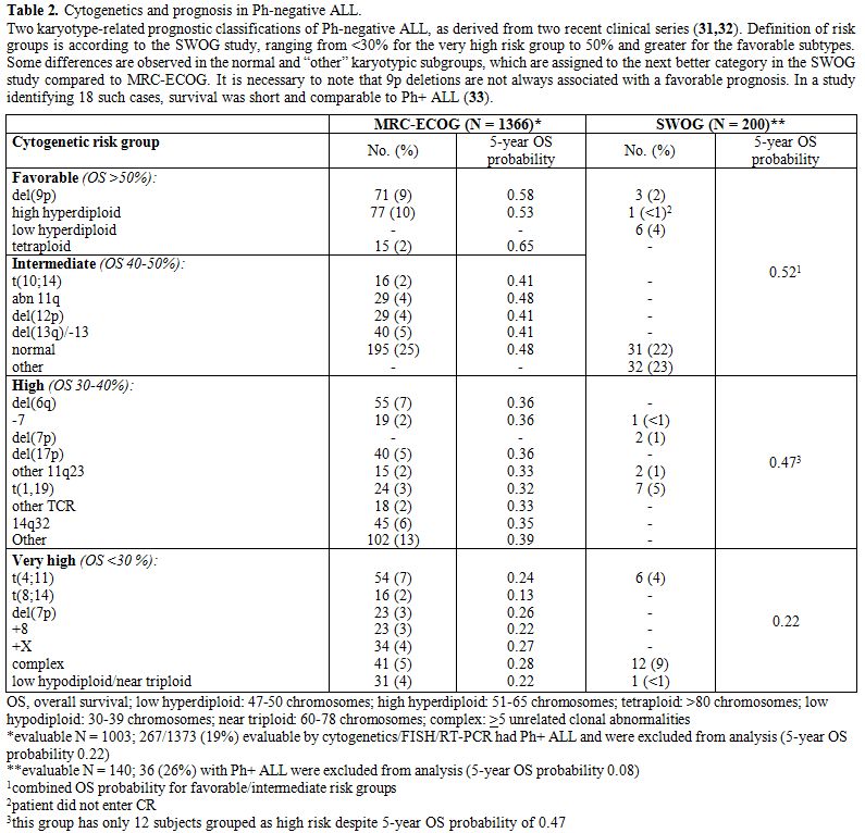
Table 2. Cytogenetics and prognosis in Ph-negative ALL.
Two karyotype-related prognostic classifications of Ph-negative ALL, as derived from two recent clinical series [31,32]. Definition of risk groups is according to the SWOG study, ranging from <30% for the very high risk group to 50% and greater for the favorable subtypes. Some differences are observed in the normal and “other” karyotypic subgroups, which are assigned to the next better category in the SWOG study compared to MRC-ECOG. It is necessary to note that 9p deletions are not always associated with a favorable prognosis. In a study identifying 18 such cases, survival was short and comparable to Ph+ ALL [33].

Cytogenetic/Genetic Risk Groups

Among the good prognosis aberrations, it is worth mentioning del(12p) or t(12p)/t(12;21)(p13;q22) in B-lineage ALL, and t(10;14)(q24;q11) in T-ALL. These abnormalities are relatively rare in adults compared with childhood ALL.
Aberrations associated with an intermediate-risk comprise the normal diploid subset plus cases with hyperdiploidy and several other recurrent or random chromosomal abnormalities.
Other aberrations, i.e. those with isolated trisomy 21, trisomy 8, and perhaps del(6q) and t(1;19)(q23;p13)/E2A-PBX1 may constitute an intermediate-high risk group; recent evidence suggests that the dismal outcome previously reported for the t(1;19)(q23;p13)/E2A-PBX1 is overcome by current therapeutic approaches.[35,36] Other recently identified aberrations in the intermediate high-risk group are represented by iAMP21[37] and IGH rearrangements, including CRLF2.[38]
Finally, patients with t(9;22)(q34;q11)or BCR-ABL1 rearrangements or a positive FISH test (Ph+ ALL), t(4;11)(q21;q23) or MLL rearrangements at 11q23, monosomy 7, hypodiploidy/low hypodiploidy (and the strictly related near triploid group) fall into the poor-risk cytogenetic category, with an overall disease-free survival (DFS) rate of about 25%, or 10% in the case of Ph+ ALL prior to the introduction of tyrosine kinase inhibitors (TKI).[39-42] Ph+ ALL may constitute 25-50% of CD10+ common or pre-B ALL cases and represent the most frequent abnormality in the adult/elderly, being detected in more than 50% of cases in 6th decade of life.[43] Secondary chromosome abnormalities in addition to t(9;22)(q34;q11) may worsen the prognosis;[44] however, this is as yet unproven in TKI era.[45] Currently, the most unfavorable group within cases with known genetic/molecular aberration is represented by t(4;11)(q21;q23) + MLL1-rearranged ALL, for which outcome is very poor unless allogeneic transplantation is adopted.[46]
Some other karyotypes are unique to specific ALL syndromes. Translocations involving chromosome 8 (MYC gene), such as t(8;14)(q24;q32) (90% of cases), t(8;22)(q24;q11)(10% of cases), and t(2;8) (rarely observed), are virtually present in 100% of cases of mature B-ALL with L3/Burkitt morphology and clonal surface immunoglobulins. Typical cytogenetic aberrations are also found in T-lineage ALL.[47] The most frequent involve 14q11 breakpoints e.g. t(10;14)(q24;q11), t(11;14)(p13;q11), or other. The presence of t(8;14) with breakpoints at q24;q11 (q24;q32 in B-ALL) in T-ALL is associated with a lymphomatous, aggressive presentation.[48,49]

New Genetics and Genomics in ALL

The integration of results of several techniques, i.e. gene expression profiling (GEP), SNP array analysis, and currently next-generation sequencing (NGS), have permitted a better definition of the molecular scenario of ALL and the identification of a constellation of novel mutations; as for the latter, however, caution must be shown, since while the biological role has been elucidated for some, while further investigation is required for others. These findings are detailed below (Tables 3, 4). 

Table 3
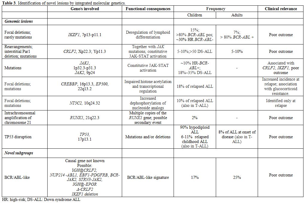
Table 3. Identification of novel lesions by integrated molecular genetics.

Table 4
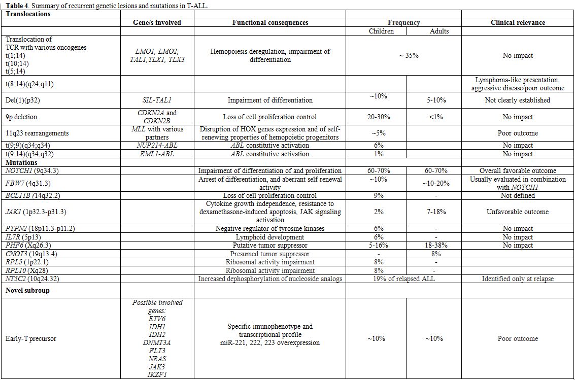
Table 4. Summary of recurrent genetic lesions and mutations in T-ALL.

B-lineage ALL: IKZF1, encoding for the transcription factor Ikaros, is frequently disrupted in BCR/ABL+ ALL (80% of cases). IKZF1 deletions, that can be different in size, are predictors of poor outcome in Ph+ ALL,[50-52] as well as in non-Ph+ ALL.[53-55]
Deregulated overexpression of CRLF2 (CRLF2), found exclusively in 5-10% B-ALL cases without known molecular rearrangements[56,57] is usually sustained by two types of aberrations: a rearrangement that involves CRLF2 and the Ig heavy chain locus (IGH@-CRLF2) or an interstitial PAR1 deletion that juxtaposes intron 1 of P2RY8 to the coding region of CRLF2 itself. More rarely, CRLF2 mutations can be detected. -CRLF2 can be detected together with IKZF1 deletion in Ph-negative ALL patients and with JAK mutations (JAK1 or JAK2) or IL7R mutations; furthermore, they are identified in roughly 50% of children with Down syndrome;[55,58] although some contrasting results have been reported, its presence correlates with an overall poor outcome.[54,55]
By the integration of genome-wide technologies, the “BCR/ABL-like” subgroup has been suggested/identified in both the adult[59,60] and pediatric populations[61,62] and it accounts for about 15% of B-ALL cases. This subgroup is characterized by a gene expression signature that is similar to that of BCR/ABL+ patients, frequent detection of IKZF1 deletions and CRLF2 rearrangements and adismal outcome. NGS has revealed the presence of mutations and/or rearrangements activating tyrosine kinases, i.e IGH-CRLF2, NUP214-ABL1 rearrangements, in-frame fusions of EBF1-PDGFRB, BCR-JAK2 or STRN3-JAK2 and cryptic IGH-EPOR rearrangements.[63] The recognition of this subgroup is of relevance, because of the poor prognosis observed. Open issues are represented by difficulty in detecting them with techniques other than gene expression profiling, which is not routinely performed in all centers, and by the fact that there is not a recurrent common lesion underlying the signature identified. With this in mind, it is plausible that the use of TKIs and/or mTOR inhibitors might be of benefit in these patients, as suggested by xenograft models.[64,65]
Hypodiploid ALL, regarded as a poor prognosis group, has been extensively evaluated in pediatric ALL:[66] NGS proved that lesions involving receptor tyrosine kinases and RAS signaling (i.e. NRAS, KRAS, FLT3 and NF1) can be detected in up to 70% of near haploid cases, whereas low hypodiploid cases are characterized by lesions involving members of the Ikaros family, particularly IKZF2, and by TP53 disruptions, that can be identified in 91.2% of these cases. In adult ALL, these cases are characterized by nonrandom chromosomal losses and the CDKN2A/B locus deletion as sole recurrent abnormality; as already reported in children, these cases frequently harbor TP53 mutations.[67]
TP53 disruption has been also recently evaluated in childhood and adult ALL. In children[68-71] this is detected in 6.4% and 11.1% of relapsed B-ALL and T-ALL cases, and, in a smaller minority of cases, also at diagnosis. A correlation with poorer outcome has been shown. In adults, TP53 mutations are identified at diagnosis in 8.2% of cases (11.1% T-ALL and 6.4% B-ALL), and are preferentially identified in cases without molecular aberrations, where they are detected in 14% of cases, and are associated with refractoriness to chemotherapy.
Other lesions identified by NGS in B-lineage ALL, are represented by mutations in CREBBP and its paralogue, EP300 (p300),[72] which were identified in the relapse samples and appear to be more frequent in hyperdiploid relapsed cases.[73] Similarly, NT5C2 mutations, which confer increased enzymatic activity on the NT5C2 protein, which normally dephosphorylates nucleoside analogs, such as mercaptopurine, used in consolidation and maintenance therapy, have been described.[74] Results are summarized in Table 3.
T-lineage ALL: In T-ALL, well-recognized aberrations include the T-cell receptor (TCR) gene rearrangements, chromosomal deletions, and focal gene deletions (Table 4).[75-83] Moreover, chromosomal rearrangements can also lead to in-frame fusion genes encoding chimeric proteins with oncogenic properties such as PICALM-MLLT10, NUP214-ABL1 fusion formed on episomes, EML-ABL1, SET-NUP214 fusion and MLL gene rearrangements with numerous different partners. The prognostic significance of these lesions is uncertain.
Furthermore, the ETP subgroup and/or myeloid-like subgroup emerged as a grey zone between AML and T-ALL by applying genome-wide technologies.[18,84,85] Initially, the reported incidence of this subgroup was established at around 10% of T-ALL cases; however, with the better recognition of these cases, its frequency is likely to be higher. Immunophenotype is characterized by an early T-cell phenotype and co-expression of at least one myeloid marker, while at the transcriptional level they have a stem-cell like profile with overexpression of myeloid transcription factors (including CEBPA, CEBPB, CEBPD), and a set of micro-RNAs (miR-221, miR-222 and miR-223). NGS has highlighted the presence of mutations usually found in acute myeloid leukemia (IDH1, IDH2, DNMT3A, FLT3 and NRAS),[86] as well mutations in the ETV6 gene. Finally, these cases rarely harbor NOTCH1 mutations.[87] Overall, prognosis is poor in these cases.
A large set of mutations (Table 4) has been identified in T-ALL by re-sequencing and NGS: they include NOTCH1, FBW7, BCL11B, JAK1, PTPN2, IL7R and PHF6, beyond those identified in ETPs; some of them have recognized prognostic significance, whereas for others further studies are required. In fact, NOTCH1 and/or FBW7 mutations, which occur in more than 60% and about 20% of cases, respectively, are usually associated with a favorable outcome. In the light of this, a prognostic model has been recently proposed, defining as low-risk patients those who harbor NOTCH1 and FBW7 mutations, and as high risk those without these mutations or with lesions involving RAS/PTEN.[83,88-91] At variance, JAK1 mutations, which increase JAK activity and alter proliferation and survival have been associated with chemotherapy refractoriness and should be considered as poor prognostic markers.[92-94]
Finally, another group of mutations/lesions is possibly involved in leukemogenesis, but their prognostic impact is either unknown or absent. They include: 1) BCL11B lesions, which can induce a developmental arrest and aberrant self-renewal activity;[95,96] 2) PTPN2 - a negative regulator of tyrosine kinases-, mutations, often detected in TLX1 overexpressing cases, T-ALL, NUP214-ABL+ patients and JAK1 mutated cases;[97,98] 3) mutations in IL7Ralpha, that lead to constitutive JAK1 and JAK3 activation and enhancement of cell cycle progression;[99,100] 4) PHF6 mutations;[101,102] 5) mutations in PTPRC, encoding the protein tyrosine phosphatase CD45, usually detected in combination with activating mutations of IL7R, JAK1 or LCK, and associated with downregulation of CD45 expression;[103] 6) mutations in CNOT3, presumed to be a tumor suppressor; 7) mutations of RPL5 and RPL10, which impair ribosomal activity.[104] Lastly, similarly to what is observed in relapsed B-ALL, NT5C2 mutations.[105]

Concluding Remarks

Due to the reviewed evidence and the complexity of all the issues at play, it is recommended that adult patients with ALL should be treated within prospective clinical trials, which is the best way to ensure both diagnostic accuracy and therapeutic efficacy. In the context of a modern risk- and subset-oriented therapy, the early diagnostic work-up is of the utmost importance and therefore needs to be carried out by well trained and highly experienced personnel (Figure 4). As a first step, it is mandatory to differentiate rapidly Ph+ from Ph-ALL and to distinguish between major immunophenotypic subsets in the latter group. The remaining diagnostic elements are available at a later stage and permit a proper identification and treatment of the several disease and risk entities. Ongoing research will permit the further definition of novel subgroups with prognostic significance.

Figure 4
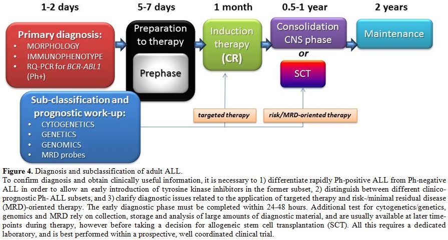
Figure 4. Diagnosis and subclassification of adult ALL.
To confirm diagnosis and obtain clinically useful information, it is necessary to 1) differentiate rapidly Ph-positive ALL from Ph-negative  ALL in order to allow an early introduction of tyrosine kinase inhibitors in the former subset, 2) distinguish between different clinico-prognostic Ph- ALL subsets, and 3) clarify diagnostic issues related to the application of targeted therapy and risk-/minimal residual disease (MRD)-oriented therapy. The early diagnostic phase must be completed within 24-48 hours. Additional test for cytogenetics/genetics, genomics and MRD rely on collection, storage and analysis of large amounts of diagnostic material, and are usually available at later time-points during therapy, however before taking a decision for allogeneic stem cell transplantation (SCT). All this requires a dedicated laboratory, and is best performed within a prospective, well coordinated clinical trial.

References

  1. Vardiman JW, Thiele J, Arber DA, et al. The 2008 revision of the World Health Organization (WHO) classification of myeloid neoplasms and acute leukemia: rationale and important changes. Blood 2009;114:937-951. http://dx.doi.org/10.1182/blood-2009-03-209262         
  2. Bennett JM, Catovsky D, Daniel MT, et al. French-American-British (FAB Cooperative Group). Proposals for the classification of the acute leukaemias. Br J Haematol 1976; 33:451-458. http://dx.doi.org/10.1111/j.1365-2141.1976.tb03563.x PMid:188440       
  3. Bennett JM, Catovsky D, Daniel MT, et al. The morphological classification of acute lymphoblastic leukaemia: concordance among observers and clinical correlations. Br J Haematol 1981; 47:553-561. http://dx.doi.org/10.1111/j.1365-2141.1981.tb02684.x PMid:6938236       
  4. Jaffe ES, Harris NL, Stein H, et al. Introduction an overview of the classification of the lymphoid neoplasms. In: Swerdlow SH, Campo E, Harris NL, et al, eds. WHO Classification of tumours of haematopoietic and lymphoid tissue. IARC: Lyon 2008;158-166. PMid:18283093       
  5. Lai R, Hirsch-Ginsberg CF, Bueso-Ramos C. Pathologic diagnosis of acute lymphocytic leukemia .Hematol Oncol Clin North Am.2000;14:1209-1235. http://dx.doi.org/10.1016/S0889-8588(05)70183-0     
  6. d'Onofrio G, Zini G, BainBJ (Translator). Morphology of Blood Disorders, 2nd Edition. Wiley-Blackwell, 2014. ISBN: 978-1-118-44260)     
  7. Sevilla DW, Colovai AI, Emmons FN, et al. Hematogones: a review and update. Leuk Lymphoma 2010;51:10-19. http://dx.doi.org/10.3109/10428190903370346   PMid:20001239       
  8. Stein P, Peiper S, Butler D, et al. Granular acute lymphoblastic leukemia. Am J Clin Pathol 1983;80:545.PMid:6578676       
  9. Gassmann W, Löffler H, Thiel E, et al. Morphological and cytochemical findings in 150 cases of T-lineage acute lymphoblastic leukaemia in adults. German Multicentre ALL Study Group (GMALL). Br J Haematol 1997; 97:372-382. http://dx.doi.org/10.1046/j.1365-2141.1997.d01-2171.x  PMid:9163604       
  10. Bene MC, Castoldi G, Knapp W, et al. Proposals for the immunological classification of acute leukemias. European Group for the Immunological Characterization of Leukemias (EGIL). Leukemia 1995;9:1783-1786. PMid:7564526       
  11. Béné MC, Nebe T, Bettelheim P, et al. Immunophenotyping of acute leukemia and lymphoproliferative disorders: a consensus proposal of the European LeukemiaNet Work Package 10. Leukemia 2011;25:567-574. http://dx.doi.org/10.1038/leu.2010.312 PMid:21252983       
  12. Kalina T, Flores-Montero J, van der Velden VH, et al. EuroFlow standardization of flow cytometer instrument settings and immunophenotyping protocols. Leukemia 2012;26:1986-2010. http://dx.doi.org/10.1038/leu.2012.122 PMid:22948490 PMCid:PMC3437409     
  13. van Dongen JJ, Lhermitte L, Böttcher S, et al. EuroFlow antibody panels for standardized n-dimensional flow cytometric immunophenotyping of normal, reactive and malignant leukocytes. Leukemia 2012;26:1908-1975. http://dx.doi.org/10.1038/leu.2012.120  PMid:22552007 PMCid:PMC3437410     
  14. Coustan-Smith E, Behm FG, Sanchez J, et al. Immunological detection of minimal residual disease in children with acute lymphoblastic leukaemia..Lancet 1998;351:550-554. http://dx.doi.org/10.1016/S0140-6736(97)10295-1     
  15. Janossy G, Coustan-Smith E, Campana D. The reliability of cytoplasmic CD3 and CD22 antigen expression in the immunodiagnosis of acute leukemia: a study of 500 cases. Leukemia 1989;3:170-181 PMid:2465463       
  16. Lenormand B, Bene MC, Lesesve JF, et al. PreB1 (CD10-) acute lymphoblastic leukemia: immunophenotypic and genomic characteristics, clinical features and outcome in 38 adults and 26 children. The Groupe dEtude Immunologique des Leucemies. Leuk Lymphoma 1998;28:329-342. PMid:9517504       
  17. Geijtenbeek TB, van Kooyk Y, van Vliet SJ, et al. High frequency of adhesion defects in B-lineage acute lymphoblastic leukemia. Blood 1999;94:754-764.  PMid:10397743       
  18. Coustan-Smith E, Mullighan CG, Onciu M, et al: Early T-cell precursor leukaemia: a subtype of very high-risk acute lymphoblastic leukaemia. Lancet Oncol 2009;10:147-156. http://dx.doi.org/10.1016/S1470-2045(08)70314-0     
  19. Vardiman JW, Thiele J, Arber DA, et al. The 2008 revision of the World Health Organization (WHO) classification of myeloid neoplasms and acute leukemia: rationale and important changes. Blood 2009;114:937-951. http://dx.doi.org/10.1182/blood-2009-03-209262 PMid:19357394       
  20. Matutes E, Pickl WF, Van't Veer M, et al. Mixed-phenotype acute leukemia: clinical and laboratory features and outcome in 100 patients defined according to the WHO 2008 classification. Blood 2011;117:3163-171. http://dx.doi.org/10.1182/blood-2010-10-314682 PMid:21228332       
  21. Paietta E, Neuberg D, Richards S, et al. Rare adult acute lymphocytic leukemia with CD56 expression in the ECOG experience shows unexpected phenotypic and genotypic heterogeneity. Am J Hematol 2001;66:189-196. http://dx.doi.org/10.1002/1096-8652(200103)66:3<189::AID-AJH1043>3.0.CO;2-A     
  22. Kantarjian HM, Hirsch-Ginsberg C, Yee G, et al. Mixed-lineage leukemia revisited: acute lymphocytic leukemia with myeloperoxidase-positive blasts by electron microscopy. Blood 1990;76:808-813. PMid:2166608       
  23. Hans CP, Finn WG, Singleton TP, et al. Usefulness of anti-CD117 in the flow cytometric analysis of acute leukemia. Am J Clin Pathol 2002;117:301-305. http://dx.doi.org/10.1309/RWCG-E5T9-GU95-LEWE PMid:11863227       
  24. Faber J, Kantarjian H, Roberts MW, Keating et al. Terminal deoxynucleotidyl transferase-negative acute lymphoblastic leukemia. Arch Pathol Lab Med 2000;124:92-97  PMid:10629138      
  25. Zhou Y, Fan X, Routbort M, et al Absence of terminal deoxynucleotidyl transferase expression identifies a subset of high-risk adult T-lymphoblastic leukemia/lymphoma.Mod Pathol 2013;26:1338-1345. http://dx.doi.org/10.1038/modpathol.2013.78 PMid:23702731       
  26. Pui CH, Crist WM, Look AT. Biology and clinical significance of cytogenetic abnormalities in childhood acute lymphoblastic leukemia. Blood 1990;76:1449-1463.   PMid:2207320      
  27. Secker-Walker LM, Prentice HG, et al. Cytogenetics adds independent prognostic information in adults with acute lymphoblastic leukaemia on MRC trial UKALL XA. MRC Adult Leukaemia Working Party. Br J Haematol 1997;96:601-610. http://dx.doi.org/10.1046/j.1365-2141.1997.d01-2053.x  PMid:9054669       
  28. Group Français de Cytogénétique Hématologique. Cytogenetic abnormalities in adult acute lymphoblastic leukemia: correlations with hematologic findings outcome. A Collaborative Study of the Group Français de Cytogénétique Hématologique.. Blood 1996;88:3135-314.     
  29. Wetzler M, Dodge RK, Mrozek K, et al. Prospective karyotype analysis in adult acute lymphoblastic leukemia: the cancer and leukemia Group B experience. Blood 1999;93:3983-3993. PMid:10339508       
  30. Kolomietz E, Al-Maghrabi J, Brennan S, et al. Primary chromosomal rearrangements of leukemia are frequently accompanied by extensive submicroscopic deletions and may lead to altered prognosis. Blood 2001;97:3581-3588. http://dx.doi.org/10.1182/blood.V97.11.3581  PMid:11369654       
  31. Moorman AV, Harrison CJ, Buck GA, et al. Karyotype is an independent prognostic factor in adult acute lymphoblastic leukemia (ALL): analysis of cytogenetic data from patients treated on the Medical Research Council (MRC) UKALLXII/Eastern Cooperative Oncology Group (ECOG) 2993 trial. Blood 2007;109:3189-3197. http://dx.doi.org/10.1182/blood-2006-10-051912 PMid:17170120       
  32. Pullarkat V, Slovak ML, Kopecky KJ, Forman SJ, Appelbaum FR. Impact of cytogenetics on the outcome of adult acute lymphoblastic leukemia: results of Southwest Oncology Group 9400 study. Blood 2008;111:2563-2572. http://dx.doi.org/10.1182/blood-2007-10-116186 PMid:18156492 PMCid:PMC2254550     
  33. Nahi H, Hagglund H, Ahlgren T, et al. An investigation into whether deletions in 9p reflect prognosis in adult precursor B-cell acute lymphoblastic leukemia: a multi-center study of 381 patients. Haematologica 2008;93:1734-1738. http://dx.doi.org/10.3324/haematol.13227 PMid:18728022       
  34. Charrin C, Thomas X, Ffrench M, et al. A report from the LALA-94 and LALA-SA groups on hypodiploidy with 30 to 39 chromosomes and near-triploidy: 2 possible expressions of a sole entity conferring poor prognosis in adult acute lymphoblastic leukemia (ALL). Blood 2004;104:2444-2451. http://dx.doi.org/10.1182/blood-2003-04-1299 PMid:15039281       
  35. Felice MS, Gallego MS, Alonso CN, et al. Prognostic impact of t(1;19)/ TCF3-PBX1 in childhood acute lymphoblastic leukemia in the context of Berlin-Frankfurt-Münster-based protocols. Leuk Lymphoma 2011;52:1215-1221. http://dx.doi.org/10.3109/10428194.2011.565436  PMid:21534874       
  36. Burmeister T, Gökbuget N, Schwartz S, Fischer L, Hubert D, Sindram A, Hoelzer D, Thiel E. Clinical features and prognostic implications of TCF3-PBX1 and ETV6-RUNX1 in adult acute lymphoblastic leukemia. Haematologica 2010;95:241-246. http://dx.doi.org/10.3324/haematol.2009.011346  PMid:19713226       PMCid:PMC2817026     
  37. Harrison CJ, Moorman AV, Schwab C, et al. An international study of intrachromosomal amplification of chromosome 21 (iAMP21): cytogenetic characterization and outcome. Leukemia 2014;28:1015-1021 http://dx.doi.org/10.1038/leu.2013.317 PMid:24166298       
  38. Moorman AV, Schwab C, Ensor HM, et al. IGH@ translocations, CRLF2 deregulation, and microdeletions in adolescents and adults with acute lymphoblastic leukemia J Clin Oncol 2012; 30:3100-3108. http://dx.doi.org/10.1200/JCO.2011.40.3907  PMid:22851563       
  39. Schardt C, Ottmann OG, Hoelzer D, Ganser A. Acute lymphoblastic leukemia with the (4;11) translocation: combined cytogenetic, immunological and molecular genetic analyses. Leukemia 1992;6:370-374. PMid:1375695       
  40. Faderl S, Albitar M. Insights into the biologic and molecular abnormalities in adult acute lymphocytic leukemia. Hematol Oncol Clin North Am. 2000;14:1267-1288. http://dx.doi.org/10.1016/S0889-8588(05)70186-6     
  41. Westbrook CA, Hooberman AL, Spino C, et al. Clinical significance of the BCR-ABL fusion gene in adult acute lymphoblastic leukemia: a Cancer and Leukemia Group B Study (8762). Blood. 1992;80:2983-2990 PMid:1467514       
  42. Gleissner B, Gökbuget N, Bartram CR, et al, German Multicenter Trials of Adult Acute Lymphoblastic Leukemia Study Group. Leading prognostic relevance of the BCR-ABL translocation in adult acute B-lineage lymphoblastic leukemia: a prospective study of the German Multicenter Trial Group and confirmed polymerase chain reaction analysis. Blood 2002;99:1536-1543. http://dx.doi.org/10.1182/blood.V99.5.1536 PMid:11861265       
  43. Chiaretti S, Vitale A, Cazzaniga G, et al.Clinico-biological features of 5202 patients with acute lymphoblastic leukemia enrolled in the Italian AIEOP and GIMEMA protocols and stratified in age cohorts. Haematologica. 2013;98:1702-1710. http://dx.doi.org/10.3324/haematol.2012.080432 PMid:23716539 PMCid:PMC3815170     
  44. Rieder H, Ludwig WD, Gassmann W, et al. Prognostic significance of additional chromosome abnormalities in adult patients with Philadelphia chromosome positive acute lymphoblastic leukaemia. Br J Haematol. 1996;95:678-691. http://dx.doi.org/10.1046/j.1365-2141.1996.d01-1968.x PMid:8982045       
  45. Ravandi F, Jorgensen JL, Thomas DA, et al. Detection of MRD may predict the outcome of patients with Philadelphia chromosome-positive ALL treated with tyrosine kinase inhibitors plus chemotherapy. Blood. 2013 Aug 15;122:1214-1221. http://dx.doi.org/10.1182/blood-2012-11-466482 PMid:23836561 PMCid:PMC3976223     
  46. Marks DI, Moorman AV, Chilton L, et al. The clinical characteristics, therapy and outcome of 85 adults with acute lymphoblastic leukemia and t(4;11)(q21;q23)/MLL-AFF1 prospectively treated in the UKALLXII/ECOG2993 trial. Haematologica. 2013;98:945-952. http://dx.doi.org/10.3324/haematol.2012.081877 PMid:23349309  PMCid:PMC3669452     
  47. Schneider NR, Carroll AJ, Shuster JJ, et al. New recurring cytogenetic abnormalities and association of blast cell karyotypes with prognosis in childhood T-cell acute lymphoblastic leukemia: a pediatric oncology group report of 343 cases. Blood. 2000;96:2543-2549. PMid:11001909       
  48. Lange BJ, Raimondi SC, Heerema N, et al. Pediatric leukemia/lymphoma with t(8;14)(q24;q11). Leukemia 1992;6:613-618. PMid:1385638     
  49. Parolini M, Mecucci C, Matteucci C, et al. Highly aggressive T-cell acute lymphoblastic leukemia with t(8;14)(q24;q11): extensive genetic characterization and achievement of early molecular remission and long-term survival in an adult patient. Blood Cancer J. 2014 Jan 17;4:e176. http://dx.doi.org/10.1038/bcj.2013.72 PMid:24442205 PMCid:PMC3913941     
  50. Martinelli G, Iacobucci I, Storlazzi CT, et al: IKZF1 (Ikaros) deletions in BCRABL1-positive acute lymphoblastic leukemia are associated with short disease-free survival and high rate of cumulative incidence of relapse: a GIMEMA AL WP report. J Clin Oncol 2009; 27:5202-5207. http://dx.doi.org/10.1200/JCO.2008.21.6408    PMid:19770381       
  51. Mullighan CG, Su X, Zhang J, et al: Children's Oncology Group: Deletion of IKZF1 and prognosis in acute lymphoblastic leukemia. N Engl J Med 2009; 360:470-480. http://dx.doi.org/10.1056/NEJMoa0808253 PMid:19129520 PMCid:PMC2674612     
  52. van der Veer A, Zaliova M, Mottadelli F, et al. IKZF1 status as a prognostic feature in BCR-ABL1-positive childhood ALL. Blood 2014; 123:1691-8. http://dx.doi.org/10.1182/blood-2013-06-509794 PMid:24366361       
  53. Kuiper RP, Waanders E, van der Velden VH, et al: IKZF1 deletions predict relapse in uniformly treated pediatric precursor B-ALL. Leukemia 2010; 24:1258-1264. http://dx.doi.org/10.1038/leu.2010.87 PMid:20445578       
  54. Moorman AV, Schwab C, Ensor HM, et al. IGH@ translocations, CRLF2 deregulation, and microdeletions in adolescents and adults with acute lymphoblastic leukemia J Clin Oncol 2012; 30:3100-3108. http://dx.doi.org/10.1200/JCO.2011.40.3907 PMid:22851563       
  55. van der Veer A, Waanders E, Pieters R, et al. Independent prognostic value of BCR-ABL1-like signature and IKZF1 deletion, but not high CRLF2 expression, in children with B-cell precursor ALL. Blood 2013;122:2622-2629. http://dx.doi.org/10.1182/blood-2012-10-462358 PMid:23974192 PMCid:PMC3795461     
  56. Mullighan CG, Collins-Underwood JR, Phillips LA, et al: Rearrangement of CRLF2 in B-progenitor- and Down syndrome-associated acute lymphoblastic leukemia. Nat Genet 2009; 41:1243-1246. http://dx.doi.org/10.1038/ng.469 PMid:19838194 PMCid:PMC2783810     
  57. Yoda A, Yoda Y, Chiaretti S, et al. Yoda A, Yoda Y, Chiaretti S, et al: Functional screening identifies CRLF2 in precursor B-cell acute lymphoblastic leukemia. Proc Natl Acad Sci U S A 2010; 107:252-257. http://dx.doi.org/10.1073/pnas.0911726107 PMid:20018760 PMCid:PMC2806782     
  58. Hertzberg L, Vendramini E, Ganmore I, et al: Down syndrome acute lymphoblastic leukemia, a highly heterogeneous disease in which aberrant expression of CRLF2 is associated with mutated JAK2: a report from the International BFM Study Group. Blood 2010; 115:1006-1117. http://dx.doi.org/10.1182/blood-2009-08-235408  PMid:19965641       
  59. Haferlach T, Kohlmann A, Schnittger S, et al: Global approach to the diagnosis of leukemia using gene expression profiling. Blood 2005; 106:1189-1198. http://dx.doi.org/10.1182/blood-2004-12-4938 PMid:15878973       
  60. Chiaretti S, Li X, Gentleman R, et al. Gene expression profiles of B-lineage adult acute lymphocytic leukemia reveal genetic patterns that identify lineage derivation and distinct mechanisms of transformation. Clin Cancer Res. 2005;11:7209-7219. http://dx.doi.org/10.1158/1078-0432.CCR-04-2165  PMid:16243790       
  61. Den Boer ML, van Slegtenhorst M, De Menezes RX, et al: A subtype of childhood acute lymphoblastic leukaemia with poor treatment outcome: a genome-wide classification study. Lancet Oncol 2009; 10:125-134. http://dx.doi.org/10.1016/S1470-2045(08)70339-5     
  62. Harvey RC, Mullighan CG, Wang X, et al: Identification of novel cluster groups in pediatric high-risk B-precursor acute lymphoblastic leukemia with gene expression profiling: correlation with genome-wide DNA copy number alterations, clinical characteristics, and outcome. Blood 2010; 116:4874-4884.  http://dx.doi.org/10.1182/blood-2009-08-239681 PMid:20699438 PMCid:PMC3321747     
  63. Roberts KG, Morin RD, Zhang J, et al. Genetic alterations activating kinase and cytokine receptor signaling in high-risk acute lymphoblastic leukemia. Cancer Cell 2012;22:153-166. http://dx.doi.org/10.1016/j.ccr.2012.06.005 PMid:22897847 PMCid:PMC3422513     
  64. Maude SL, Tasian SK, Vincent T, et al. Targeting JAK1/2 and mTOR in murine xenograft models of Ph-like acute lymphoblastic leukemia. Blood 2012;120:3510-3518. http://dx.doi.org/10.1182/blood-2012-03-415448 PMid:22955920  PMCid:PMC3482861     
  65. Roberts KG, Li Y, Payne-Turner D, et al. Targetable kinase-activating lesions in Ph-like acute lymphoblastic leukemia. N Engl J Med 2014;371:1005-1015.   PMid:25207766       
  66. Holmfeldt L, Wei L, Diaz-Flores E, et al. The genomic landscape of hypodiploid acute lymphoblastic leukemia. Nat Genet 2013;45:242-252. http://dx.doi.org/10.1038/ng.2532 PMid:23334668 PMCid:PMC3919793     
  67.  Mühlbacher V, Zenger M, Schnittger S, et al. Acute lymphoblastic leukemia with low hypodiploid/near triploid karyotype is a specific clinical entity and exhibits a very high TP53 mutation frequency of 93%. Genes Chromosomes Cancer 2014;53:524-536. http://dx.doi.org/10.1002/gcc.22163 PMid:24619868       
  68. Hof J, Krentz S, van Schewick, et al: Mutations and deletions of the TP53 gene predict nonresponse to treatment and poor outcome in first relapse of childhood acute lymphoblastic leukemia. J Clin Oncol 2011; 29:3185-93. http://dx.doi.org/10.1200/JCO.2011.34.8144 PMid:21747090       
  69. Krentz S, Hof J, Mendioroz A, et al: Prognostic value of genetic alterations in children with first bone marrow relapse of childhood B-cell precursor acute lymphoblastic leukemia. Leukemia 2013;27:295-304. http://dx.doi.org/10.1038/leu.2012.155 PMid:22699455       
  70. Chiaretti S, Brugnoletti F, Tavolaro S, et al: TP53 mutations are frequent in adult acute lymphoblastic leukemia cases negative for recurrent fusion genes and correlate with poor response to induction therapy. Haematologica 2013;98:e59-61. http://dx.doi.org/10.3324/haematol.2012.076786 PMid:23403321 PMCid:PMC3640132     
  71. Stengel A, Schnittger S, Weissmann S, et al. TP53 mutations occur in 15.7% of ALL and are associated with MYC-rearrangement, low hypodiploidy and a poor prognosis. Blood 2014;124:251-258. http://dx.doi.org/10.1182/blood-2014-02-558833 PMid:24829203       
  72. Mullighan CG, Zhang J, Kasper LH, et al: CREBBP mutations in relapsed acute lymphoblastic leukaemia. Nature 2011; 471:235-239. http://dx.doi.org/10.1038/nature09727 PMid:21390130 PMCid:PMC3076610     
  73. Inthal A, Zeitlhofer P, Zeginigg M, et al. CREBB HAT domain mutations prevail in relapse cases of high hyperdiploid childhood acute lymphoblastic leukemia. Leukemia. 2012;26:1797-1803. http://dx.doi.org/10.1038/leu.2012.60 PMid:22388726 PMCid:PMC4194312     
  74. Meyer JA, Wang J, Hogan LE, et al. Relapse-specific mutations in NT5C2 in childhood acute lymphoblastic leukemia. Nat Genet 2013; 45:290-294. http://dx.doi.org/10.1038/ng.2558  PMid:23377183 PMCid:PMC3681285     
  75. Barber KE, Martineau M, Harewood L, et al. Amplification of the ABL gene in T-cell acute lymphoblastic leukemia. Leukemia 2004;18:1153-1156. http://dx.doi.org/10.1038/sj.leu.2403357 PMid:15057249       
  76. Ferrando AA, Neuberg DS, Dodge RK, et al. Prognostic importance of TLX1 (HOX11) oncogene expression in adults with T-cell acute lymphoblastic leukaemia. Lancet 2004;363:535-536. http://dx.doi.org/10.1016/S0140-6736(04)15542-6     
  77. Ballerini P, Busson M, Fasola S, et al. NUP214-ABL1 amplification in t(5;14)/HOX11L2-positive ALL present with several forms and may have a prognostic significance. Leukemia 2005;19:468-470. http://dx.doi.org/10.1038/sj.leu.2403654 PMid:15674415       
  78. Asnafi V, Buzyn A, Thomas X, et al. Impact of TCR status and genotype on outcome in adult T-cell acute lymphoblastic leukemia: a LALA-94 study. Blood 2005;105:3072-3078. http://dx.doi.org/10.1182/blood-2004-09-3666 PMid:15637138       
  79. Burmeister T, Gokbuget N, Reinhardt R, Rieder H, Hoelzer D, Schwartz S. NUP214-ABL1 in adult T-ALL: the GMALL study group experience. Blood 2006;108:3556-3559. http://dx.doi.org/10.1182/blood-2006-04-014514 PMid:16873673       
  80. Baldus CD, Burmeister T, Martus P, et al. High expression of the ETS transcription factor ERG predicts adverse outcome in acute T-lymphoblastic leukemia in adults. J Clin Oncol. 2006;24:4714-4720. http://dx.doi.org/10.1200/JCO.2006.06.1580 PMid:16954520       
  81. Baldus CD, Martus P, Burmeister T, et al. Low ERG and BAALC expression identifies a new subgroup of adult acute T-lymphoblastic leukemia with a highly favorable outcome. J Clin Oncol. 2007;25:3739-3745. http://dx.doi.org/10.1200/JCO.2007.11.5253 PMid:17646667       
  82. Bergeron J, Clappier E, Radford I, et al. Prognostic and oncogenic relevance of TLX1/HOX11 expression level in T-ALLs. Blood. 2007;110:2324-2330. http://dx.doi.org/10.1182/blood-2007-04-079988 PMid:17609427       
  83. Asnafi V, Buzyn A, Le Noir S, et al: NOTCH1/FBXW7 mutation identifies a large subgroup with favourable outcome in adult T-cell acute lymphoblastic leukemia (TALL): a GRAALL study. Blood 2009; 113:3918-3924 http://dx.doi.org/10.1182/blood-2008-10-184069 PMid:19109228       
  84. Chiaretti S, Messina M, Tavolaro S, et al: Gene expression profiling identifies a subset of adult T-cell acute lymphoblastic leukemia with myeloid-like gene features and over-expression of miR-223. Haematologica 2010; 95:1114-1121. http://dx.doi.org/10.3324/haematol.2009.015099 PMid:20418243 PMCid:PMC2895035     
  85. Coskun E, Neumann M, Schlee C, et al. MicroRNA profiling reveals aberrant microRNA expression in adult ETP-ALL and functional studies implicate a role for miR-222 in acute leukemia. Leuk Res 2013;37:647-56. http://dx.doi.org/10.1016/j.leukres.2013.02.019 PMid:23522449       
  86. Zhang J, Ding L, Holmfeldt L, Wu G, et al: The genetic basis of early T-cell precursor acute lymphoblastic leukaemia. Nature 2012; 481:157-163. http://dx.doi.org/10.1038/nature10725 PMid:22237106 PMCid:PMC3267575     
  87. Van Vlierberghe P, Ambesi-Impiombato A, Perez-Garcia A, et al: ETV6 mutations in early immature human T cell leukemias. J Exp Med 2011; 208:2571-2579. http://dx.doi.org/10.1084/jem.20112239 PMid:22162831 PMCid:PMC3244026     
  88. Park MJ, Tak T, Oda M, et al: FBXW7 and NOTCH1 mutations in childhood T cell acute lymphoblastic leukaemia and T cell non-Hodgkin lymphoma. Br J Haematol 2009; 145:198-206. http://dx.doi.org/10.1111/j.1365-2141.2009.07607.x  PMid:19245433       
  89. Mansour MR, Sulis ML, Duke V, et al: Prognostic implications of NOTCH1 and FBXW7 mutations in adults with T-cell acute lymphoblastic leukemia treated on the MRC UKALLXII/ECOG E2993 protocol. J Clin Oncol 2009; 27:4352-4356. http://dx.doi.org/10.1200/JCO.2009.22.0996 PMid:19635999 PMCid:PMC2744275     
  90. Ben Abdelali R, Asnafi V, Leguay T, et al: Group for Research on Adult Acute Lymphoblastic Leukemia: Pediatric-inspired intensified therapy of adult T-ALL reveals the favorable outcome of NOTCH1/FBXW7 mutations, but not of low ERG/BAALC expression: a GRAALL study. Blood 2011; 118:5099-5107. http://dx.doi.org/10.1182/blood-2011-02-334219 PMid:21835957       
  91. Trinquand A, Tanguy-Schmidt A, Ben Abdelali R, et al. Toward a NOTCH1/FBXW7/RAS/PTEN-based oncogenetic risk classification of adult T-cell acute lymphoblastic leukemia: a Group for Research in Adult Acute Lymphoblastic Leukemia study. J Clin Oncol. 2013;31:4333-4342. http://dx.doi.org/10.1200/JCO.2012.48.5292 PMid:24166518       
  92. Flex E, Petrangeli V, Stella L, et al: Somatically acquired Jak1 mutation in adult acute lymphoblastic leukemia. J Exp Med 2008;205:751-758. http://dx.doi.org/10.1084/jem.20072182 PMid:18362173  PMCid:PMC2292215     
  93. Jeong EG, Kim MS, Nam HK, et al: Somatic mutations of JAK1 and JAK3 in acute leukemias and solid cancers. Clin Cancer Res 2008; 14:3716-3721. http://dx.doi.org/10.1158/1078-0432.CCR-07-4839 PMid:18559588       
  94. Asnafi V, Le Noir S, Lhermitte L, et al: JAK1 mutations are not frequent events in adult T-ALL: a GRAALL study. Br J Haematol 2010;148:178-179. http://dx.doi.org/10.1111/j.1365-2141.2009.07912.x  PMid:19764985       
  95. Gutierrez A, Kentsis A, Sanda T, et al: The BCL11B tumor suppressor is mutated across the major molecular subtypes of T-cell acute lymphoblastic leukemia. Blood 2011; 118:4169-4173. http://dx.doi.org/10.1182/blood-2010-11-318873 PMid:21878675 PMCid:PMC3204734     
  96. Kraszewska MD, Dawidowska M, Szczepanski T, et al: T-cell acute lymphoblastic leukaemia: recent molecular biology findings. Br J Haematol 2012; 156:303-315. http://dx.doi.org/10.1111/j.1365-2141.2011.08957.x  PMid:22145858       
  97. Kleppe M, Lahortiga I, El Chaar T, et al: Deletion of the protein tyrosine phosphatase gene PTPN2 in T-cell acute lymphoblastic leukemia. Nat Gen 2010; 42:530-535. http://dx.doi.org/10.1038/ng.587 PMid:20473312 PMCid:PMC2957655     
  98. Kleppe M, Soulier J, Asnafi V, et al: PTPN2 negatively regulates oncogenic JAK1 in T-cell acute lymphoblastic leukemia. Blood 2011; 117:7090-7098. http://dx.doi.org/10.1182/blood-2010-10-314286 PMid:21551237      
  99. Shochat C, Tal N, Bandapalli OR, et al. Gain-of-function mutations in interleukin-7 receptor-a (IL7R) in childhood acute lymphoblastic leukemias. J Exp Med. 2011;208:901-908. http://dx.doi.org/10.1084/jem.20110580 PMid:21536738 PMCid:PMC3092356     
  100. Zenatti PP, Ribeiro D, Li W, et al: Oncogenic IL7R gain-of-function mutations in childhood T-cell acute lymphoblastic leukemia. Nat Genet 2011; 43:932-939. http://dx.doi.org/10.1038/ng.924 PMid:21892159           
  101. Van Vlierberghe P, Palomero T, Khiabanian H, et al: PHF6 mutations in T-cell acute lymphoblastic leukemia.Nat Genet 2010; 42:338-342 http://dx.doi.org/10.1038/ng.542 PMid:20228800 PMCid:PMC2847364     
  102. Wang Q, Qiu H, Jiang H, et al: Mutations of PHF6 are associated with mutations of NOTCH1, JAK1 and rearrangement of SET-NUP214 in T-cell acute lymphoblastic leukemia. Haematologica 2011; 96:1808-1814. http://dx.doi.org/10.3324/haematol.2011.043083 PMid:21880637 PMCid:PMC3232263    
  103. Porcu M, Kleppe M, Gianfelici V, et al: Mutation of the receptor tyrosine phosphatase PTPRC (CD45) in T-cell acute lymphoblastic leukemia. Blood 2012; 119:4476-4479. http://dx.doi.org/10.1182/blood-2011-09-379958 PMid:22438252      
  104. De Keersmaecker K, Atak ZK, Li N, et al: Exome sequencing identifies mutation in CNOT3 and ribosomal genes RPL5 and RPL10 in T-cell acute lymphoblastic leukemia. Nat Genet 2013;45:186-190. http://dx.doi.org/10.1038/ng.2508 PMid:23263491       
  105. Tzoneva G, Perez-Garcia A, Carpenter Z, et al: Activating mutations in the NT5C2 nucleotidase gene drive chemotherapy resistance in relapsed ALL. Nat Med 2013;19:368-371. http://dx.doi.org/10.1038/nm.3078 PMid:23377281 PMCid:PMC3594483     

[TOP]