Impact of Mannose-Binding Protein Gene Polymorphisms in Omani Sickle Cell Disease Patients

Mathew Zachariah1, Shoaib Al Zadjali2, Wafa Bashir1, Rahma Al Ambusaidi2, Rhea Misquith2, Yasser Wali1 and Anil Pathare2

1 Departments of Child Health, Sultan Qaboos University Hospital, Muscat, Oman.
2 Departments of Haematology, Sultan Qaboos University Hospital, Muscat, Oman.

Corresponding author: Dr. Anil Pathare, MD, FCPS, FIMSA, Ph.D. Sultan Qaboos University Hospital, PO Box 35, PC 123, Muscat, OMAN. E-mail: pathare@squ.edu.om 

Published: February 15, 2016
Received: October 9, 2015
Accepted: Jamuary 11, 2016
Mediterr J Hematol Infect Dis 2016, 8(1): e2016013, DOI 10.4084/MJHID.2016.013
This article is available on PDF format at:

This is an Open Access article distributed under the terms of the Creative Commons Attribution License
(
https://creativecommons.org/licenses/by-nc/4.0), which permits unrestricted use, distribution, and reproduction in any medium, provided the original work is properly cited.

Abstract

Objectives: Our aim was to study mannose-binding protein (MBP) polymorphisms in exonic and promoter region and correlate it with associated infections and vasoocculsive (VOC) episodes in sickle cell disease (SCD) patients since MBP plays an important role in innate immunity by activating the complement system.
Methods: We studied the genetic polymorphisms in the Exon 1 (alleles A/O) and promoter region (alleles Y/X; H/L, P/Q) of the MBL2 gene, in SCD patients as an increased incidence of infections is seen in these patients. A PCR-based, targeted genomic DNA sequencing of MBL2 was used to study 68 SCD Omani patients and 44 controls (healthy voluntary blood donors).
Results: In SCD patients, the frequency of the genotype related to the high production of MBL was 0.35 (YA/YA) and for intermediate/low production was 0.65 (YA/XA, XA/XA, YA/YO, XA/YO, YO/YO). The observed frequencies of MBL2 gene promoter polymorphism (-221, Y/X) were 44.4% and 20.5% for the heterozygous genotype Y/X and 3.2% and 2.2% for the homozygous (X/X) respectively between SCD patients and controls. MBL2 Exon1 gene mutations were 29.4% and 50% for the heterozygous genotype A/O and 5.9% and 6.8% respectively for the homozygous (O/O) genotype between SCD patients and controls. The distribution of variant MBL2 gene polymorphisms did not show any correlation in SCD patients with or without VOC attacks (p=0.16; OR-0.486; CI=0.177-1.33), however, it was correlated with infections (p=0.0162; OR-3.55; CI 1.25-10.04).
Conclusions: Although the frequency of the genotypes and haplotypes of MBL2 in SCD patients did not differ from controls, overall in the SCD patient cohort the increased representation of variant alleles was significantly correlated with infections (p<0.05). However, these variant MBL2 polymorphisms did not seem to play a significant role in the VOC episodes in this SCD cohort.


Introduction

Sickle cell disease (SCD) is characterized by a striking variability in the clinical presentation ranging from an early-onset life-threatening disease to a milder condition compatible with an almost normal life course. Polymerization of deoxy Hb S with red blood cell (RBC) deformation, desiccation and increased rigidity results in painful vasoocclusive crises (VOC) and hemolytic anemia. Adherence of sickle RBC stimulates endothelial cells to upregulate their adhesion molecules, which accelerates the adhesion cascade.[1] Activated endothelium also releases a broad range of cytokines, including granulocyte-macrophage-colony-stimulating factor (GM-CSF), interleukin (IL)-1, IL-3, IL-6, IL-8 and tumor necrosis factor (TNF-a), and these have been detected in the plasma of patients with SCD.[2,3] Neutrophils may also become activated during this cascade of vasoocclusive events, and neutrophil adherence may contribute to vasoocclusion,[3] as well as endothelial cell damage.[4]
Patients with SCD have an increased tendency to infection,[5] especially with encapsulated organisms, which is due in part to the poor splenic function,[6] but might also be a feature of altered neutrophil and monocyte function.[7] There is now ample evidence indicative of an ongoing inflammatory state between painful crises in SCD involving neutrophils, monocyte activation and an abnormality of cytokine-regulated neutrophil function, which may compromise the host defenses against certain microorganisms.[8] In this context, polymorphism of the mannose-binding lectin (MBL) has been documented as potential immunogenetic modulating factors that could constitute an additional risk of infection in SCD.[9] However, polymorphisms of the Fc receptor may, in fact, protect SCD patients from infections with H. influenzae.[10]
Mannose-binding lectin (MBL) is a serum protein of hepatic origin belonging to a family of Ca2+-dependent collagenous lectins, most of which are components of the innate immune system or natural immunity.[11,12] Mutations in the mannose binding protein gene have been associated with recurrent infections.[13-15] A single gene, MBL2, located on chromosome 10, codes for human MBL.[16,17] Mannose-binding lectin may exert its action through binding to mannose-rich, and N-acetyl-glucosamine oligosaccharides present on a variety of microorganisms. Therefore, it activates the complement system by MBL-associated serine proteases and by interacting with novel receptors on phagocytes.[18-20]
The mannose binding protein, being part of the innate immune system, is considered particularly important in the vulnerable period of infancy before an adequate specific immune protection was attained by the adaptive immune system.[21]
Five single-nucleotide polymorphisms influencing serum MBL levels have been identified.[22] Three variant alleles have been described in exon 1 of the MBL2 gene.[23-25] These variants are due to 3 single-base pair substitutions at codon 54 (allele B), codon 57 (allele C), and codon 52 (allele D). They, independently, cause low serum MBL levels.[23] The normal wild type allele is commonly designated A, and the three mutant alleles O. All variant alleles reduce the amount of functional MBL subunits in heterozygous individuals 5-to 10-fold.[26] The serum MBL concentration is also dependent on some nucleotide substitutions in the promoter region of the MBL2 gene.[27,28] In particular, a polymorphism in codon -221 (X/Y type) has a significant effect on the MBL serum concentration with the Y promoter having high and the X having low MBL-expressing activity. [26-28]
Given the relatively high prevalence of SCD in the Omani population,[29] we decided to study the genetic polymorphism of MBL in children and adolescent patients with SCD. The aim of the study is to establish the MBL genotypes in the SCD patients as well as in the Omani ethnic population. The study also attempted clinical correlation with the type, severity of infections and complication seen in SCD patients like VOC’s.

Materials and Methods

Patients: The study was conducted on 68 Omani SCD patients, aged between 3–18 years (mean age ± SD; 9.4± 3.9; M:F 37:31), who were enrolled into this case-control study. They were treated at the Pediatric Hematology Unit, Department of Child Health, Sultan Qaboos University Hospital (SQUH). Forty-four ethnically matched healthy voluntary blood donors were included in this study as a control group. The healthy controls were ethnic Omani subjects who were volunteer blood donors aged between 21–44 years (mean age ± SD; 26.4± 2.5; M:F 31:13). They were screened with a CBC and HPLC to confirm that they were indeed normal and then their DNA was extracted for MBP polymorphism study. This study was undertaken after approval by the institutional research and ethics committee and written informed consents were obtained from the patients, guardians in case of minor patients and controls before enrollment. A thorough history and comprehensive examination were conducted with particular emphasis on infections, history of vasooclusive crises and SCD complications. The diagnosis of SCD was confirmed by molecular studies in all the patients to characterize the SCD subtypes and haplotypes. Amongst the 68 SCD patients, 56 patients (82%) were HbSS, 11(17%) were Sickle Thal double heterozygotes, and 1(1%) was Sickle HbD [SD] double heterozygote. All the clinical and laboratory details were obtained from the electronic medical file records in all patients. Patients with infection symptoms were all investigated to document a microbiologically blood culture/ urine culture proven infection.
DNA studies: A 5ml blood sample was collected in tubes containing EDTA. Genomic DNA was isolated using the semi-automated ABI PRISM™ 6100 Nucleic Acid Prep Station, [Applied Biosystems, Foster City, CA, USA] and samples were stored at -20°C pending analysis. All the DNA polymorphisms were studied by direct sequencing of the relevant PCR- amplified genome segment on ABI PRISM™ 3100 Genetic Analyzer (Applied Biosystems, Foster City, CA, USA).
Genotyping of Exon 1 of MBL2 gene: Genotyping of exon1 (codon 52, 54 and 57 for alleles D, B, C respectively) was done by multiplex PCR as previously described.[30]
Genotyping of the promoter region of MBL2 gene: Genotyping of the promoter region was performed by direct sequencing of the corresponding region of interest (-65 for P/Q alleles, -221 for X/Y alleles and -618 for H/L alleles) by appropriate primers.[31]
Analysis of haplotypes and genotypes:  Haplotypes of MBL2 gene were divided into three groups according to Garrett et al.[32]
Statistical Methods: Data was analyzed using STATA ver. 11.1 (StataCorp, College Station, TX, USA). Numerical data were expressed as mean, standard deviation, range. Qualitative data were expressed as frequency and percentage. Chi-square was used to study the statistical significance of qualitative variables. Fisher’s exact test was used with Yates correction wherever applicable. Odds ratio [OR] and 95% confidence intervals [CI] were calculated for risk estimation. A two-sided p value of less than 0.05 was considered as statistically significant. The observed and expected genotype frequencies were analyzed by using weighted least square estimates of allele frequencies and chi-square goodness-of-fit test to see if Hardy-Weinberg’s proportions were respected.

Results

The study initially had recruited 85 SCD patients and 50 voluntary blood donor controls. However, as DNA results were available only in 68 SCD patients and 44 normal controls, this was the study population analyzed in further details and reported herein. There were 37 males and 31 females in the patient group. Their age ranged from 3 to 18 years with the mean age of 9.4 years.
The results of genotype and allele frequencies of the Exon-1 and promoter MBL2 gene polymorphisms are shown in Table 1. MBP exon1 and promoter variants were grouped into haplotypes, and all the possible groupings of the observed alleles A/O (B, C, D), H/L, Y/X and P/Q, combined, homozygote and heterozygote, with SCD and no SCD were analyzed and tabulated. In the patients with SCD the MBL2 exon-1 allele was significantly higher while the promoter alleles Q and Y were lower than in controls, (p <0.05). Furthermore, the combined genotype YA/YO was also down represented in the SCD affected, whereas, the minor mutant alleles (O, X and Q) did not obey Hardy Weinberg’s rule.


Table 1 
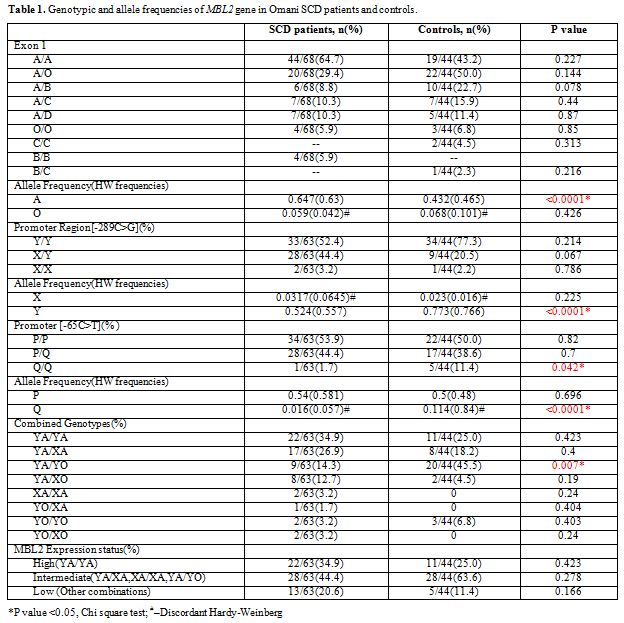
Table 1. Genotypic and allele frequencies of MBL2 gene in Omani SCD patients and controls.

SCD patients were classified into three categories based on the clinical severity index according to our previous studies.[33,34] Specifically, sickle cell disease patients were categorized as having a mild, moderate, or severe systemic disease based on the history of admissions/year for VOC events and associated clinical SCD complications like Acute chest syndrome, Osteonecrosis, Splenic sequestration, Stroke, Priapism and repeated infections. Patients with more than three hospital admissions/year and/or SCD complications named above were severe cases whereas those with less than one hospital admission for VOC/year were mild, and those, needing between 1-3 admissions/year and  SCD complications, were moderate cases.
Table 2 shows the distribution of MBL2 exon-1 A allele and the mutant allele O and significance of the correlation between the MBL2 polymorphisms and presence or absence of VOC or infections in this cohort of SCD patients. The homozygous or heterozygous mutants [OO+AO] were less common in SCD patients with or without VOC, but this difference was not statistically significant. However, they were more frequent in SCD patients with infections, and the difference was statistically significant (p<0.05).
Of 25 patients (37%), suspected to have an associated infection, 11(44%) had a microbiologically had blood or urine documented culture. In this group of 11 SCD patients, 6 had positive blood cultures (3-Gram positive cocci, and one each with Bacillus spp., E.coli and Achromobacter spp.) The remaining 5 SCD patients had positive urine cultures (3-Klebsiella pneumoniae and 2-E. coli).


Table 2 
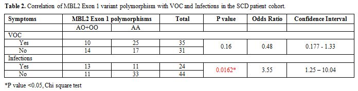
Table 2. Correlation of MBL2 Exon 1 variant polymorphism with VOC and Infections in the SCD patient cohort.


Discussion

The MBL2 gene is highly polymorphic and shows allelic variations in different ethnic groups.[25] MBL, the product of MBL2 gene is an important component of innate immunity, and functionally significant mutations decrease serum MBL levels.[18,19] In the current study, MBL2 allelic frequencies were not significantly different in the SCD group as compared to the control subjects, especially the clinically relevant MBL2 expression status (high, intermediate and low; Table 1). Although small differences were observed in the Egyptian and Brazilian SCD patients, in both these studies, like our study, there were no statistically significant allelic differences seen between controls and SCD patients.[35,36]
Our study, however, found an evident association between MBL2 variants with infection with an odds ratio of 3.55 (Table 2). This observation is in accordance with several other previous studies.[9,11]  Neonato et al. (1999) reported that mutations in MBL2, which were associated with low levels of MBL, showed increased susceptibility to infections in their cohort of SCD patients.[9] MBL is probably involved in the recognition of sickle RBC’s and removing them from the site of inflammation.[9] However with low MBL levels in patients with mutant alleles; this process is hampered. Thus, although MBL is known to be associated with innate immunity, the presence of variant MBL2 genes is considered to affect the efficiency of complement-phagocyte interaction leading to an increased propensity for infections, especially of encapsulated microorganisms as suggested by increased pneumococcal infections.[37] This interpretation is also corroborated by the routine administration of the pneumococcal vaccine in SCD children below five years inducing a decrease of pneumococcal infections.[38] Unfortunately, in this study we were unable to measure the serum MBL levels, although it was planned as part of this study, due to certain technical shortcomings.
Curiously, a couple of studies have reported the association of MBL variant genes with vasooclusive crisis from Brazilian SCD patients. Oliveira et al. (2009) and Mendonca et al. (2010) reported that there was an association between MBL2 structural polymorphism with VOC.[36,39] In our study, we did not find any association between MBL2 variants with VOC (Table 2). This difference is due to the characterization of VOC events, which was quite different. In both these studies from Brazil, the presence of only one VOC was used as an index of SCD severity, whereas, in our study we have classified patients as severe with three or more than three VOC events/year, and mild with one event.[33,34] This classification is recommended by the Cooperative Study of Sickle Cell Disease (CSSCD).[40]
Furthermore, SCD patients from Oman are a mixture of several African and Asian βs haplotypes with 52.1% showing Benin haplotypes, 26.7% showing Arab-Indian haplotypes, and 21.4% showing Bantu haplotypes.[41] It is thus apparent that almost two-thirds of these patients have a severe form of disease and hence have a higher severity index.[33,34] VOC’s are a complex process with an interaction between sickle reticulocytes, perturbed endothelium, and inflammatory cytokines. It is thus hard to mechanistically understand how such complex interactions could be affected by MBL2 polymorphisms which as stated above are well correlated to susceptibility to infections.
Oman has shown a remarkable progress in several fields like education, health care and socio-economic progress in the last several decades. However, autosomal recessive disorders like SCD persist in the population due to the practice of consanguineous marriages.[42] Thus, there is quite a disparity in the observed and expected frequency of minor MBL2 alleles X, O, and Q where the Hardy-Weinberg equation is not obeyed (Table 1). Nevertheless, it is certain that MBL2 variant alleles are significantly associated with infections in SCD patients from Oman.

Acknowledgments

We wish to thank the Hospital Administration for allowing the use of hospital data. This work was supported by an internal grant (IG/MED/CHILD/06/01) from the College of Medicine and Health Sciences.

References

  1. Natarajan M, Udden MM, McIntire LV: Adhesion of sickle red blood cells and damage to interleukin-1 stimulated endothelial cells under flow in vitro. Blood , 1996,  87(11): 4845-52. PMID: 8639858.    
  2. Croziat H: Circulating cytokines in sickle cell patients during steady state. Br J Haematol,  1994,87(3): 592-7. PMID: 7527647    
  3. Pathare AV, Salam Al Kindi, S Daar, and David Dennison, “Cytokines in Sickle Cell Disease”, Hematology, 2003, 8 (5), 329-337. PMID: 14530175. http://dx.doi.org/10.1080/10245330310001604719    
  4. Varani J, Ginsburg I, Schuger L, et al: Endothelial cell killing by neutrophils: Synergistic interaction of oxygen products and proteases. Am J Pathol, 1989, 135(3): 435-8. PMID: 2675621    
  5. Francis RB, Johnson CS: Vascular occlusion in sickle cell disease: Current concepts and unanswered questions. Blood 1991, 77(7): 1405-14. PMID: 2009364.    
  6. Leone G, Pizzigallo E. Bacterial Infections Following Splenectomy for Malignant and Nonmalignant Hematologic Diseases. Mediterr J Hematol Infect Dis. 2015 ;7(1):e2015057. doi: 10.4084/MJHID.2015.057. eCollection 2015. Review.    
  7. Boghossian SH, Wright G, Webster DB, Segal AW: Investigations of host defence in patients with sickle cell disease. Br J Haematol, 1985, 59(3): 523-31. PMID: 3882140.    
  8. Mollapour E, Porter JB, Kaczmarski R., Linch DC, and Roberts P.J.: Raised neutrophil phospholipase A2 activity and defective priming of NADPH oxidase and phospholipase A2 in Sickle cell disease. Blood, 1998, 91(9): 3423-9. PMID: 9558401.    
  9. Neonato, M.G., Lu, C.Y., Guilloud-Bataille, M., et al., Genetic polymorphism of the mannose-binding protein gene in children with sickle cell disease: identification of three new variant alleles and relationship to infections. Eur J Hum Genet,1999 ,7(6): 679-86. http://dx.doi.org/10.1038/sj.ejhg.5200360    
  10. Norris, CF, Surrey, S, Bunin, G.R.,  et al , Relationship between Fc receptor IIA polymorphism and infection in children with sickle cell disease. J Pediatr 1996, 128 (6), 813-9. http://dx.doi.org/10.1016/S0022-3476(96)70334-7    
  11. Turner MW. Mannose-binding lectin: the pluripotent molecule of the innate immune system. Immunol Today. 1996,;17(11): 532-540. http://dx.doi.org/10.1016/S0167-5699(96)80908-X     
  12. Sumiya M, Summerfield JA. Mannose-binding protein, genetic variants and the risk of infection. Q J Med 1996;89(10): 723-6. http://dx.doi.org/10.1093/qjmed/89.10.723    
  13. Sumiya M, Super M, Tabona P, et al. Molecular basis of opsonic defect in immunodeficient children. Lancet. 1991; 337(8757):, 1569-1570. http://dx.doi.org/10.1016/0140-6736(91)93263-9    
  14. Garred P, Madsen HO, Hofmann B, Svejgaard A. Increased frequency of homozygosity of abnormal mannan-binding-protein alleles in patients with suspected immunodeficiency. Lancet. 1995,346 (8980): 941-943. http://dx.doi.org/10.1016/S0140-6736(95)91559-1    
  15. Summerfield JA, Ryder S, Sumiya M, et al. Mannose binding protein gene mutations associated with unusual and severe infections in adults. Lancet. ;345 (8954): 1995, 886-889. http://dx.doi.org/ 10.1016/S0140-6736(95)90009-8    
  16. Sastry K, Herman GA, Day L, et al. The human mannose-binding protein gene. J Exp Med. 1989, ;170 (4): 1175-1189. PMID: 2477486 http://dx.doi.org/10.1084/jem.170.4.1175     
  17. Taylor ME, Brickell PM, Craig RK, Summerfield JA. Structure and evolutionary origin of the gene encoding a human serum mannose-binding protein. Biochem J. 1989,;262 (3): 763-771. http://dx.doi.org/10.1042/bj2620763    
  18. Matsushita M, Fujita T. Activation of the classical complement pathway by mannose-binding protein in association with a novel C1s-like serine protease. J Exp Med. 1992,  ;176 (6): 1497-1502. http://dx.doi.org/10.1084/jem.176.6.1497    
  19. Thiel S, Vorup-Jensen T, Stover CM, et al. A second serine protease associated with mannan-binding lectin that activates complement. Nature. 1997; 386(6624):, 506-510. http://dx.doi.org/10.1038/386506a0    
  20. Nepomuceno RR, Henschen-Edman AH, Burgess WH, Tenner AJ. cDNA cloning and primary structure analysis of C1qR(P), the human C1q/MBL/SPA receptor that mediates enhanced phagocytosis in vitro. Immunity. 1997,;6 (2): 119-129. http://dx.doi.org/10.1016/S1074-7613(00)80419-7    
  21. Turner MW. Mannose binding protein. Biochem Soc Trans. 1994;22(1):, 88-94. http://dx.doi.org/10.1042/bst0220088     
  22. Turner MW. Mannose-binding lectin (MBL) in health and disease. Immunobiology. 1998;199 (2):, 327-339. http://dx.doi.org/10.1016/S0171-2985(98)80037-5    
  23. Lipscombe RJ, Sumiya M, Hill AV, et al. High frequencies in African and non-African populations of independent mutations in the mannose binding protein gene. Hum Mol Genet. 1992,;1 (9): 709-715. http://dx.doi.org/10.1093/hmg/1.9.709    
  24. Madsen HO, Garred P, Kurtzhals JA, et al. A new frequent allele is the missing link in the structural polymorphism of the human mannan-binding protein. Immunogenetics. 1994 ;40 (1):, 37-44. PMID: 8206524.    
  25. Garred P, Madsen HO, Svejgaard A. Genetics of human mannan-binding protein. In: Ezekowitz RAB, Sastry K, Reid KBM, eds. Collectins and Innate Immunity. Austin, Tex: RG Landes; 1996, 139-164.    
  26. Madsen HO, Garred P, Thiel S, et al. Interplay between promotor and structural gene variants control basal serum level of mannan-binding protein. J Immunol. 1995,;155 (6): 3013-3020. PMID: 7673719    
  27. Madsen HO, Satz ML, Høgh B, et al. Different molecular events result in low protein levels of mannan-binding lectin in populations from southeast Africa and South America. J Immunol. 1998,;161 (6): 3169-3175. PMID: 9743385    
  28. Super M, Thiel S, Lu J, Levinsky RJ, Turner MW. Association of low levels of mannan-binding protein with a common defect of opsonisation. Lancet. 1989;2 (8674): 1236-1239. http://dx.doi.org/10.1016/S0140-6736(89)91849-7    
  29. Al Kindi S; Zadjali S; Al Madhani A; et al, Forcasting hemoglobinopathy burden through neonatal screening in Omani neonates,  Hemoglobin.; 2010, 34(2):135-44. http://dx.doi.org/10.3109/03630261003677213    
  30. Skalnikova H, Freibergera T, Chumchalova J, Grombinkova H, Sediva A,  Cost-effective genotyping of human MBL2 gene mutations using multiplex PCR. J. Immunol Methods, 2004, 295(1-2),139-147. http://dx.doi.org/10.1016/j.jim.2004.10.007    
  31. Sullivan KE, Wooten C, Goldman D, Petri M Mannose binding protein genetic polymorphisms in black patients with systemic lupus erythromatosus. Arthritis Rheum, 1996:39(12):2046-2051. PMID: 8961910    
  32. Garred P, Mannose-binding lectin genetics: from A to Z. Biochem Soc. Trans,  2008, 36(Pt6):1461-1466. http://dx.doi.org/10.1042/BST0361461    
  33. Wali YA, Al-Lamki Z, Hussein SS, et al. Splenic function in Omani children with sickle cell disease: correlation with severity index, hemoglobin phenotype, iron status, and alpha-thalassemia trait. Pediatr Hematol Oncol.2002 ;19 (7):491S-500. http://dx.doi.org/10.1080/08880010290097314    
  34. Ganesh A, Al-Zuhaibi S, Pathare A, et al. Orbital infarction in sickle cell disease. Am J Ophthalmol.2008;146 (4):595-601.    http://dx.doi.org/10.1016/j.ajo.2008.05.041        
  35. El Deen MAK, Khorsheid MM, El Sadani ZA, Amrousy YM, Galal NM, Mannose-binding lectin (MBL2) gene polymorphism in sickle cell anemia : an Egytptian study, Comp. Clin. Pathol,  epub. 26th Jan 2012. http://dx.doi.org/10.1007/s00580-012-1420-y    
  36. Mendonça TF, Oliveira MC, Vasconcelos LR, et al. Association of variant alleles of MBL2 gene with vasoocclusive crisis in children with sickle cell anemia. Blood Cells Mol Dis.2010 ; 44(4):224-8. http://dx.doi.org/10.1016/j.bcmd.2010.02.004     
  37. Jack DL, Dodds AW, Anwar N, et al. Activation of complement by mannose-binding lectin on isogenic mutants of Neisseria meningitidis serogroup B. J Immunol.1998 ;160(3):1346-53. PMID: 9570553    
  38. Halasa NB, Shankar SM, Talbot TR, et al. Incidence of invasive pneumococcal disease among individuals with sickle cell disease before and after the introduction of the pneumococcal conjugate vaccine. Clin Infect Dis.; 2007, 44(11):1428-33. http://dx.doi.org/10.1086/516781     
  39. Oliveira MC, Mendonça TF, Vasconcelos LR, et al. Association of the MBL2 gene EXON1 polymorphism and vasoocclusive crisis in patients with sickle cell anemia. Acta Haematol.2009 ;121(4):212-5. http://dx.doi.org/10.1159/000220335    
  40. Gaston M, Smith J, Gallagher D, et al. Recruitment in the Cooperative Study of Sickle Cell Disease (CSSCD) Control Clin Trials. 1987 Dec;8(4 Suppl):131S-140S.    
  41. Daar S, Hussain HM, Gravell D, Nagel RL, Krishnamoorthy R. Genetic epidemiology of HbS in Oman: multicentric origin for the betaS gene. Am J Hematol.2000 ;64(1):39-46. http://dx.doi.org/10.1002/(SICI)1096-8652(200005)64:1<39::AID-AJH7>3.0.CO;2-#        
  42. Rajab A, Patton MA. A study of consanguinity in the Sultanate of Oman. Ann Hum Biol.2000, 27(3):321-6. PMID: 10834296.    

[TOP]