Identifying High-Risk Chronic Lymphocytic Leukemia: A Pathogenesis-Oriented Appraisal of Prognostic and Predictive Factors in Patients Treated with Chemotherapy with or without Immunotherapy

Sara Martinelli, Antonio Cuneo, Luca Formigaro, Maurizio Cavallari, Enrico Lista, Francesca Maria Quaglia, Maria Ciccone, Antonella Bardi, Eleonora Volta, Elisa Tammiso, Elena Saccenti, Olga Sofritti, Giulia Daghia, Massimo Negrini, Melissa Dabusti, Paolo Tomasi, Sabrina Moretti, Francesco Cavazzini and Gian Matteo Rigolin

Section of Hematology, Azienda Ospedaliero-Universitaria S.Anna, Ferrara, Italy.

Corresponding author: Antonio Cuneo. Section of Hematology, Azienda Ospedaliero-Universitaria S.Anna. Via Aldo Moro 8, 44124, Cona, Ferrara, Italy. Tel.: +39-0532-236977, Fax.: +39-0532-236654. E-mail: cut@unife.it

Published: October 15, 2016
Received: December 2, 2015
Accepted: September 16,2016
Mediterr J Hematol Infect Dis 2016, 8(1): e2016047, DOI 10.4084/MJHID.2016.047
This article is available on PDF format at:

This is an Open Access article distributed under the terms of the Creative Commons Attribution License
(http://creativecommons.org/licenses/by/2.0), which permits unrestricted use, distribution, and reproduction in any medium, provided the original work is properly cited.


Abstract

Chronic lymphocytic leukemia (CLL) displays an extremely variable clinical behaviour. Accurate prognostication and prediction of response to treatment are important in an era of effective first-line regimens and novel molecules for high risk patients. Because a plethora of prognostic biomarkers were identified, but few of them were validated by multivariable analysis in comprehensive prospective studies, we applied in this survey stringent criteria to select papers from the literature in order to identify the most reproducible prognostic/predictive markers. Each biomarker was analysed in terms of reproducibility across the different studies with respect to its impact on time to first treatment (TTFT), progression free survival (PFS), overall survival (OS) and response to treatment. We were able to identify the following biomarkers as the most reliable in guiding risk stratification in the daily clinical practice: 17p-/TP53 mutations, IGHV unmutated configuration, short telomeres and 11q-. However, the method for measuring telomere length was not validated yet and 11q- was predictive of inferior OS only in those patients who did not receive FCR-like combinations. Stage and lymphocytosis were predictive of shorter TTFT and age, high serum thymidine kinase levels and poor performance status were predictive of shorter OS. Using our criteria no parameter was found to independently predict for inferior response to treatment.

Introduction

Chronic lymphocytic leukemia (CLL) displays a variable clinical behaviour, with many patients living for years without symptoms and other patients requiring early therapeutic intervention attaining short lasting responses and succumbing to their disease in a few years. Therefore, survival in this chronic lymphoproliferative disorder largely depends on the rapidity of disease progression and on the quality and duration of response to treatment. The availability of effective first-line regimens[1-6] makes prognostication and prediction of response to treatment an important exercise in clinical practice, especially in young and/or fit patients who may benefit of aggressive regimens including allogeneic bone marrow transplantation.[7,8] Clinical staging is a simple measure of disease burden and still represents a convenient, yet insufficient means of assessing prognosis, because it does not identify those patients with limited disease who have a high probability to progress, and it does not predict the quality and duration of response to treatment. A plethora of biomarkers have been identified in the last decades which may predict disease outcome,[9] but few of them were validated in the context of prospective studies using adequate statistic considerations to weigh the risk of each parameter by multivariable analysis. Meanwhile, our understanding of CLL biology greatly improved providing a basis for a better understanding of the biologic role of prognostic markers.[10] The pathogenesis of CLL is the result of a complex interplay between i) lymphocytes carrying a restricted repertoire of BCR,[11] ii) the mutational status of the variable portion of the immunoglobulin heavy chain (IGHV) gene determining different behaviour of neoplastic lymphocyte in response to antigen stimulation, iii) cell activation and interaction with the microenvironment,[12] iv) genetic lesions[13] (Figure 1). Each of these fundamental mechanisms is associated with specific biomarkers that identify high-risk CLL, as summarized in Figure 2. Central to this categorization of prognostic markers is the concept that antigenic stimulation of neoplastic lymphocytes with a restricted set of BCR may promote inhibition of apoptosis, survival and proliferation within the lymph node and bone marrow microenvironment, with consequent multiple cycles of cell division, telomere shortening and genetic instability, with disease-host interactions ultimately shaping variable clinical phenotypes (Figure 2).[14]

In this review clinicobiologic features predicting outcome are discussed in correlation with their pathogenic role and applicability in clinical practice.


Figure 1 
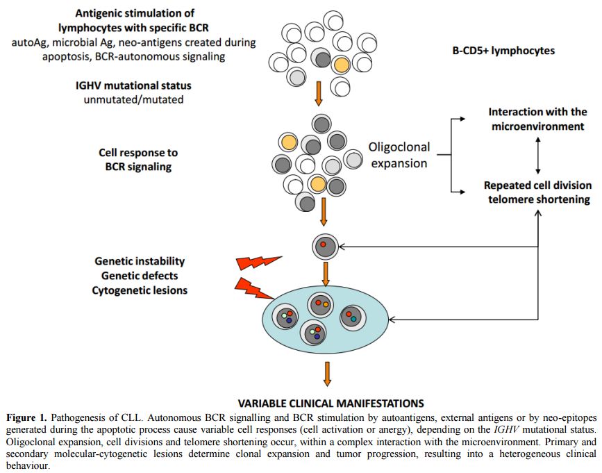
Figure 1. Pathogenesis of CLL. Autonomous BCR signalling and BCR stimulation by autoantigens, external antigens or by neo-epitopes generated during the apoptotic process cause variable cell responses (cell activation or anergy), depending on the IGHV mutational status. Oligoclonal expansion, cell divisions and telomere shortening occur, within a complex interaction with the microenvironment. Primary and secondary molecular-cytogenetic lesions determine clonal expansion and tumor progression, resulting into a heterogeneous clinical behaviour.

Figure 2
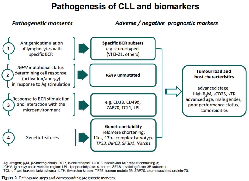
Figure 2. Pathogenic steps and corresponding prognostic markers.


Eligibility Criteria and Literature Search

Based on previous analyses that identified clinical and biologic characteristics having prognostic significance,[9,10,15-17] the following 18 biomarkers were included in this survey: stereotyped receptors and BCR subsets, CD38, ZAP70, CD49d, IGHV gene mutational status, 17p-/TP53 mutations, 11q-, telomere length, complex karyotype, NOTCH1 and SF3B1 mutations, age, gender, performance status, stage, lymphocytosis, beta-2-microglobulin, thymidine kinase.

A literature search was then performed to identify studies on the prognostic value of these biomarkers in CLL. We searched PubMed to identify all citations from January 2000 to April 2016 describing the role of the selected parameters in predicting the outcome for newly diagnosed CLL patients. The search was performed using a combination of MeSH controlled vocabulary and text words. The following terms were used: “Leukemia, Lymphocytic, Chronic, B-Cell"[Mesh], "Prognosis"[Mesh], "Clinical Trial" [Publication Type], "Receptors, Antigen, B-Cell"[Mesh], CD38, ZAP70, CD49d, IGHV, IGVH, 17p[All Fields], TP53[All Fields], 11q[All Fields], "Telomere"[Mesh], telomere, complex karyotype, NOTCH1, SF3B1, "beta 2-Microglobulin"[Mesh], thymidine kinase.

Only full length publications satisfying the following requirements were included in the review: i) English language; ii) at least 100 patients included; iii) multivariate analysis including salient clinical data and genetic testing (IGHV mutational status, 17p deletion and 11q deletion); iv) prospective design of the study (clinical trial) or single/multicentre study using a learning cohort and a validation cohort or consecutive series; v) at least one endpoint being time to first treatment (TTFT),  progression free survival (PFS), overall survival (OS), overall response rate (ORR) or complete response (CR) rate. Manuscripts describing the prognostic impact of the selected parameters in the context of patients starting unconventional or experimental treatment were not included, as well as studies including patients with monoclonal B-cell lymphocytosis.

The search criteria identified 3,845 citations. After duplicate removal and evaluation of all remaining manuscripts, 27 papers met the criteria for inclusion in this study. The characteristics and salient data of these papers are presented in Table 1.   


Table 1 
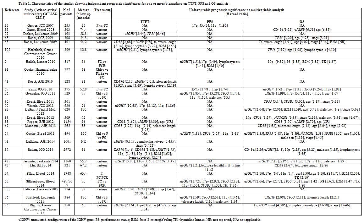
Table 1. Characteristics of the studies showing independent prognostic significance for one or more biomarkers on TTFT, PFS and OS analysis. 


Results

Predictors of outcome (TTFT, PFS and OS). Figure 3 represents, for each parameter, the total number of studies analyzing its prognostic impact and the number of studies in which the parameter showed independent prognostic significance in terms of TTFT, PFS and/or OS. Hazard ratios for each marker are reported in Table 1.


Figure 3
Figure 3. Number of studies assessing the impact of each parameter in terms of a) TTFT; b) PFS; c) OS: the blue part of each column represents the number of studies showing independent negative impact on prognosis (“significant”); the red part represents the number ofstudies showing no prognostic impact (“not significant”).

1) BCR repertoire and stereotyped receptors
Pathogenic role: CLL lymphocytes express a restricted set of BCR due to non-random usage of gene families coding for the variable portion of the Ig.[11] Furthermore, some CLL cases express highly homogeneous sequences of the heavy chain complementarity determining region 3 (HCDR3), a phenomenon referred to as “stereotyped” BCR[18] that was shown to occur in up to one-third of the cases.[19-21]
The similarity of the BCR from various CLL patients suggests that the precursors of B-CLL cells were chosen for their antigen-binding capabilities by antigen(s) of restricted nature and structure.  
Prognostic impact: Biased BCR usage and stereotyped receptors did not meet the criteria adopted in this review. The prognostic significance of stereotyped BCR was only assessed in studies that did not include a comprehensive assessment of salient genetic parameters, and no prospective clinical trial was designed including determination of BCR stereotypy in the diagnostic workup. CLL with stereotyped BCR showed shorter TTFT in a study including genetic parameters;[22] however no multivariable analysis was performed in this analysis.

2) Interaction with the microenvironment and activation markers
Pathogenic role: The interaction between neoplastic lymphocytes responding to BCR stimulation and the microenvironment plays a fundamental role in CLL pathogenesis.[23] As a consequence, the natural history of the disease is dictated, in part, by lymphocyte survival and growth in the lymph nodes that favours the accumulation of genomic alterations, especially within proliferation centres and/or CD38-positive cells.[24,25] Biomarkers reflecting the capability to respond to BCR engagement or to adhere to niches in the bone marrow or lymph node (i.e. CD38, ZAP70, CD49d, lipoprotein lipase, TCL1 expression) were correlated with progressive disease[26-30] and, to the contrary, a signature of anergic lymphocyte that poorly responds to BCR stimulation was observed in indolent CLL.[31,32]
Prognostic impact: The threshold for CD38-positivity varied across studies. In a comprehensive analysis of 1154 early stage CLL seen at 4 European centres,[33] CD38+ was predictive of a shorter TTFT (median 9.3 years vs not reached) and shorter survival (median 14,7 years) by multivariable analysis (Table 1). Interestingly a more rapid disease progression requiring treatment was observed in CD38 positive CLL in patients with “favourable” genetic profile.[25] However, the prognostic significance of CD38 was overcome by more robust genetic parameters in several clinical trials, as shown in Figure 3.[34-36]
ZAP-70 was predictive of a shorter TTFT in only one study,[37] and no impact was shown on PFS or OS in all studies using a complete assessment of prognostic parameters when the multivariable analysis was applied.[35,38-43] One study showed an independent impact of ZAP70 on OS;[44] however, it was not considered in this survey as it used an unusually low expression level (>10%) as a positivity cut-off and because the detection method is not reproducible.[9]
CD49d expression was predictive of shorter interval between diagnosis and disease progression, with a median TTFT of approximately 4 years in CD49d-positive cases.[38,44] Its predictive value was confirmed by multivariable analysis in two analyses[37,41] and an independent adverse impact on OS was documented in two studies using heterogeneous treatment.[37,39]
No study with comprehensive assessment of prognostic markers was performed to assess the independent prognostic impact of lipoproteine lipase and TCL1 overexpression. 

3) IGHV mutational status and genetic features
Pathogenic role: Neoplastic lymphocytes carrying ≤2% mutations of the IGHV gene compared to the nearest germline sequence are referred to as “unmutated” CLL. The lymphocytes in this CLL subset respond to antigen stimulation by activating intracellular signalling and entering the G1 phase of the cell cycle more efficiently than IGHV “mutated” CLL.[23,45,46] Consequently neoplastic lymphocyte carrying unmutated IGHV sequence, i) undergo more cell divisions in vivo as shown by incorporation of deuterated water[12] and, ii) carry shorter telomeres and accumulate more genomic defects[47] than lymphocytes with mutated IGHV.
The molecular pathogenesis of CLL is centred around some lesions. i.e. TCL1 or miR-29 overexpression and/or miR15a-16 deletion, producing the disease in the animal model.[48-50] These lesions are associated with a number of chromosomes and genetic aberrations which may appear soon during clonal expansion (13q-, +12, MYD88, NOTCH1 mutations), or later following disease progression or selection by treatment (11q-, 17p-, mutations of TP53, ATM, SF3B1, BIRC3).[51,52] Disruption of the TP53 and ATM pathway is associated with resistance to DNA damaging agents and genetic instability leading to the emergence of subclones accounting for disease progression.[9]
Prognostic impact:
-    Unmutated IGHV sequences: Ever since the first reports,[26,53] unmutated IGHV proved to be a robust unfavourable prognostic marker.[16] In a prospective study by Shanafelt and coworkers[38] TTFT in 1004 CLL was 2,8 years in unmutated CLL as compared to 11 years in mutated CLL. In our analysis, a significantly shorter TTFT with very high hazard ratios (Table 1) was observed in 8 prospective series, 4 of which had enrolled more than 700 cases. PFS was shorter in IGHV unmutated CLL in 2 large studies using fludarabine and cyclofosfamide (FC) or fludarabine, cyclofosfamide and rituximab (FCR)2 or chlorambucil (Chlor), fludarabine (F) or FC[36] and the observed difference held when including assessment of new gene mutations (i.e. SF3B1 and NOTCH1) and telomere length in the multivariable model.[54-56] In the CLL4 trial of the GCLLSG using F or FC the IGHV configuration in 294 patients showed no independent impact on PFS[57] as was the case with a US Intergroup Phase III Trial E2997 that assessed the IGHV mutational status in 195 patients;[35] however these 2 analyses were numerically smaller than the other studies. Interestingly, a shorter survival was noted in IGHV unmutated CLL in a number of large studies (Table 1, Figure 3) with 85,1% five-year OS in unmutated CLL vs 91.4% five-year OS in mutated CLL in a recent analysis that pooled data form 3 randomized studies of the GCLLSG.[34]
-    Telomeres: Telomeres are specific non-coding nucleotide sequences consisting of 6–12 kbp of TTAGGG-repeats located at the ends of eukaryotic chromosomes that are necessary for the complete replication and stability of the chromosome. Because they are eroded upon each cell division, their length reflects the replicative history of a cell.[58] In CLL cells telomeres are shorter that in normal B-lymphocytes[59] and those patients with telomere length below the median observed value were found to frequently carry unmutated IGHV gene[60] and unfavourable prognosis.[61] In our analysis, a significantly shorter TTFT was found in CLL with shortened telomeres in 3 studies,[41,62,63] 2 of which had partially overlapping cohorts (Table 1). Short telomeres were independently associated with inferior PFS in two large studies assessing the most significant prognostic parameters[43,56] and negatively impacted on OS in 4 studies that included patients treated with various regimens(Table 1, Figure 3).[43,56,62,63]
-    17p-/TP53 mutations and 11q deletion: These aberrations alter cell-cycle and DNA-repair pathways. In the case of a functional TP53 pathway, DNA damage activates p53 through the activation of ATM, thus inducing cell cycle arrest through p21 and apoptosis. 17p13 deletion causes loss of one TP53 allele and determines resistance to DNA-damaging drugs through haploinsufficiency.[64] 17p13 deletion is associated in the vast majority of cases with inactivating mutations of the remaining TP53 allele. The TP53 mutation may occur independent of 17p deletion and may inactivate p53 function by a dominant negative effect or by duplication of the chromosome segment containing the mutated TP53 gene, a genetic rearrangement referred to as uniparental disomy.[65] Disruption of the TP53 pathway by deletion and/or mutation leads to resistance to apoptosis and genetic instability and indeed 17p- patients frequently exhibit complex chromosome defects and multiple genetic lesions.[55,66] The negative impact of 17p13 deletion on OS and clinical response to fludarabine were clearly documented as early as 1995,[67] and a similar negative impact was shown to be associated with TP53 mutations, independent of the presence of 17p13 deletion.[36,57,68] Interestingly, minor clones carrying TP53 mutations (<20% of the cells) were equally shown by sensitive next generation sequencing techniques to predict for an inferior outcome.[69] The papers selected for this review show that the aberrations leading to disruption of the TP53 pathway, either by 17p deletion or by inactivating TP53 gene mutation, or both, have a deleterious impact on all outcome measure, as shown in Table 1 and Figure 3. A hazard ratio of 3,96 of being treated after 26 months vs. patients with 13q- as single aberration was recorded in a large prospective study of 930 patients[70] and more rapid disease progression requiring treatment was observed in one large multicentre study[71] and in one single centre study.[72] At variance, one large analysis conducted at a referral laboratory receiving samples from several institutions did not find an impact on TTFT for TP53 mutations.[42] Even though the presence of mutated IGHV gene and early stage reduced the adverse effect of this genetic aberration in a minority of patients,[73,74] 17p-/TP53 mutations definitely identify patients with dismal outcome with the current treatment regimens using alkylating agents, purine analogues, and anti CD20 monoclonal antibodies. Indeed shorter PFS and OS were uniformly reported in all the studies (Table 1, Figure 3). Interestingly, TP53 disruption appears to be associated with more frequent progression in the relapsed/refractory setting under novel active BCR-targeted therapies.[75-77] 11q22-23 deletion characterizes a CLL subtype with extensive nodal involvement and inferior prognosis.[78] The minimal region of deletion involves the ataxia-teleangiectasia mutated (ATM) gene, with concurrent mutation of the remaining ATM allele occurring in 30-40% of 11q- cases and causing extreme telomere shortening[79] and dismal prognosis.[80] Because ATM is a very large gene, mutational studies were not performed in clinical trials and papers included in this review refer to patients with 11q-. TTFT and PFS were shorter in patients with 11q- in the majority of studies (Table 1, Figure 3). It is noteworthy, however, that the negative impact on PFS was overcome by adding rituximab to FC.[2] Likewise, some reports demonstrated an adverse impact of 11q- on OS,[81] whereas more studies using effective regimens based on purine analogues and alkylating agents with rituximab[2,55,82] did not detect any difference by multivariable analysis. Thus, it appears that the negative prognostic impact of 11q- is abated in young and/or fit patients treated by modern chemoimmunotherapy regimens.
-    Lesions involving NOTCH1 and SF3B1: Mutations causing activation of the NOTCH1 pathway, with consequent activation of non-canonical NFkB signalling, may promote cell survival and resistance to apoptosis.[83-85] SF3B1 mutations cause altered splicing of a number of targets including FOXP1, that encodes for a forkhead transcription machinery,[86] promoting resistance to fludarabine-based treatment through as yet unknown mechanisms.[87] Shorter TTFT was noted in patients with SF3B1 mutations in 2 studies,[42,88] whereas NOTCH1 mutation did not predict for more rapid disease evolution (Table 1). Likewise, NOTCH1 mutations did not impact on PFS, whereas shorter PFS was observed in SF3B1 mutated patients treated by FC or FCR in the CLL8 study.[55] This correlation was not observed in the UKCLL4 study using chlorambucil, or fludarabine with or without cyclophosphamide (Table 1).[54] Rossi et al. showed an adverse impact on OS of NOTCH1 mutation.[89] OS was shorter in SF3B1 and NOTCH1 mutated patients in the UKCLL4 trial[54] and not in the CLL8 trial (Table 1).[55] It is noteworthy that two more studies showed an independent impact of the SF3B1 mutation on overall survival (Figure 3).[42,90]
-    Conventional banding analysis: Metaphase karyotyping represented the first biomarker having prognostic significance in CLL, in the seminal paper by Juliusson et al..[91] More recently complex karyotype was included in a prognostic scoring system predictive of time to first treatment and overall survival.[92] The presence of chromosome aberrations was predictive of an inferior outcome in those patients without detectable aberrations by fluorescence in situ hybridization (FISH).[93] Unbalanced chromosome translocations, frequently occurring in the context of complex karyotype, were shown to be independent prognostic factors in a study[94] and complex karyotype predicted for a shorter TTFT[71] and OS[95] by multivariable analysis. The prognostic value of complex karyotype was also demonstrated in relapsed/refractory CLL patients treated with ibrutinib-based regimens, where this parameter showed a stronger impact on the outcome than del(17p).[96] After data for this review had been collected, a comprehensive prospective study on 161 CLL patients with relevant comorbidity showed the independent prognostic role of complex karyotype for survival following chlorambucil-based chemoimmunotherapy.[97] Thus, there is mounting evidence that chromosome banding analysis with novel efficient mitogens may substantially contribute to the identification of CLL patients with adverse prognosis.

4) Disease/ host characteristics
Pathogenic role: Markers of tumor burden and proliferative activity of neoplastic cells may have an obvious impact on prognosis. Clinical stage, peripheral lymphocytosis, bone marrow infiltration pattern, serum markers such as soluble CD23 and ß2-microglobulin, an extracellular protein associated with the class I major histocompatibility complex, represented for many years valuable prognostic markers.[98] Likewise, markers of proliferative activity, i.e. lymphocyte doubling time and serum thymidine kinase-1 (TK1), a cellular enzyme involved in the DNA synthesis in the G1/S phase of the cell cycle and reflecting the number of dividing neoplastic cells, were shown to have a significant prognostic impact.
Age and gender may have an evident impact on prognosis. According to the global health observatory data repository of the WHO (available at the link http://www.who.int/gho/mortality_burden_disease/life_tables/situation_trends/en/) life expectancy in 2012 at the age of 60 was 19 and 23 years in Europe for males and females respectively (21 and 24 years in the Americas). Given the availability of treatments that are able to modify the natural history of the disease and to prolong survival,[99] host characteristics such as performance status and comorbidities may also have a significant role as prognosticators.[34,55]
Prognostic impact: The standardization of chemiluminescence immunoassay for the assessment of serum thymidine kinase-1 (TK) levels significantly facilitated the introduction of this marker into clinical practice,[100] and age-related normal reference values were recently defined and validated.[101] When this test was included among diagnostic workup within prospective trials, raised TK levels (i.e. ≥10 U/L) were predictive of shorter PFS[55] and survival in the GCLLSG trials.[34] Likewise, serum beta-2-microglobulin levels proved an independent prognostic parameter on more outcome measures in several studies (Table 1, Figure 3).
Lymphocytosis, ECOG performance status, stage and gender showed a variable impact on prognosis. Direct quantitation of the disease burden such lymphocytosis and stage are predictive of shorter TTFT and have a variable impact on other outcome measures,[102] however, they lacked significant prognostic value in the largest analysis based on pooled data from several GCLLSG studies.[34]
Age and PS had no impact on TTFT and PFS, whereas they showed significant impact on OS (Table 1, Figure 3).

Factors predictive of response to treatment
No paper with the stringent characteristics adopted in our analysis was found to address the impact of biomarkers on predicting response treatment, with two notable exceptions:

a) 17p-/TP53 mutations and 11q-.
There is evidence that carrying 17p-/TP53 mutation conveys a low probability to achieve a clinical response with chemoimmunotherapy.[67] ORR was lower in 17p-/TP53 mutated patients in 3 large randomized trials comparing FC vs FCR,[2] F vs FC[57] and Chlorambucil vs F vs FC.[81] Even though these data were not validated by multivariable analysis, the probability to attain a meaningful response was very low for the 17p- group in the CLL8 trial, with a CR rate of 0% and 5% and an ORR rate of 51,6% and 75% with FC and FCR, respectively.[2,55] Interestingly, TP53 mutations were shown to represent an independent predictive factor of shorter time to chemorefractoriness.[68]

b) Lack of efficacy of anti CD20 monoclonal antibodies rituximab and ofatumumab in NOTCH1 mutated patients.
Recent evidence was provided that response to chemoimmunotherapy containing anti-CD20 monoclonal antibodies may be negatively influenced by the presence of NOTCH1 mutations. The adjunct of ofatumumab to chlorambucil significantly improved PFS in the total population of a phase III study;[103] while not influencing response to treatment, NOTCH1 mutation was associated with shorter PFS in the ofatumamuab plus chlorambucil arm (17.7 months vs. 23.3, HR 1.86 p=0.01), making the addition of the monoclonal antibody to chlorambucil irrelevant in terms of PFS over chlorambucil alone.[103] In the CLL8 study[55] there was no significant difference in terms of ORR in the FC arm and in the FCR arm depending on the NOTCH1 mutation status; however, while the association of rituximab with FC improved ORR in patients with wild type NOTCH1 (88,1 vs. 96,6%; p<0.01), no difference in ORR was noted in NOTCH1 mutated patients in the FC and FCR arms (87,1% vs. 90%). PFS was superimposable in NOTCH1 mutated patients in the FCR (34,2 months) and FC arm (33,9 months). Taken together these data suggest that NOTCH1 mutation is a predictive marker for reduced benefit from the addition of rituximab or ofatumumab to chemotherapy. Interestingly, CLL cases carrying NOTCH1 mutations are characterized by low CD20 expression levels deriving at least in part from histone deacetylase-dependent transcriptional repression, an observation that may explain the low sensitivity of these patients to anti-CD20 monoclonal antibodies.[104]

Conclusion

Using stringent criteria we were able to identify 16 parameters, i.e. CD38, CD49d, unmutated IGHV, 17p-/TP53 mutations, 11q-, telomere length, complex karyotype, NOTCH1 and SF3B1 mutations, age, gender, performance status (PS), stage, lymphocytosis, beta-2-microglobulin, thymidine kinase, with unfavourable prognostic significance on TTFT, PFS and/or OS by multivariable analysis in prospective clinical trials or in the context of well-organized studies.  The observed TTFT, PFS, and OS for each of these markers in the corresponding studies is shown in Figure 4.  


Figure 4
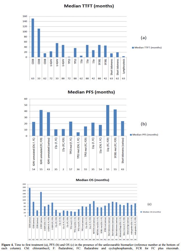
Figure 4. Time to first treatment (a), PFS (b) and OS (c) in the presence of the unfavourable biomarker (reference number at the bottom ofeach column). Chl: chlorambucil; F: fludarabine; FC: fludarabine and cyclophosphamide, FCR for FC plus rituximab.

Since 17p-/TP53 mutations, unmutated IGHV gene configuration and 11q- proved to be independent predictors of outcome in at least 2/3 studies (Figure 3) and their detection methods were standardized,[105-107] these markers are to be considered the most reliable for usage in clinical practice. Telomere length proved a reproducible predictor of the outcome but the detection method is not standardized yet, even if a recent study confirmed the reproducibility of results obtained with monochrome multiplex Q-PCR (MMQ-PCR) and single telomere length analysis (STELA), opening the way for the assay standardisation.[56] Complex karyotype as detected by stimulation with novel mitogens[66] may represent a novel predictor of unfavourable outcome.[71,95-97]
While simple measures of disease burden such as stage and lymphocytosis do not play a role any more as prognostic markers, host characteristics such as poor PS and advanced age still play a relevant role in predicting OS.
It is noteworthy that evolution of treatment may overcome the significance of some of these prognostic factors; thus, while maintaining its predictive value on OS in patients not eligible to modern  chemoimmunotherapy regimens, 11q- lost its unfavourable significance in those patients treated by FCR.[34,55] Importantly, mechanism-based treatment in the refractory/relapsed setting showed high efficacy in “high risk” patients;[75] thus the introduction of new oral agents targeting kinase signalling or BCL2 will likely change the significance and role of many of these markers.[108]

References

  1. Keating MJ, O'Brien S, Albitar M, Lerner S, Plunkett W, Giles F, et al. 2005. Early results of a chemoimmunotherapy regimen of fludarabine, cyclophosphamide, and rituximab as initial therapy for chronic lymphocytic leukemia. J Clin Oncol. 23:4079-88. http://dx.doi.org/10.1200/JCO.2005.12.051 PMid:15767648      
  2. Hallek M, Fischer K, Fingerle-Rowson G, Fink AM, Busch R, Mayer J, et al. 2010. Addition of rituximab to fludarabine and cyclophosphamide in patients with chronic lymphocytic leukaemia: a randomised, open-label, phase 3 trial. Lancet. 376:1164-74. http://dx.doi.org/10.1016/S0140-6736(10)61381-5     
  3. Fischer K, Cramer P, Busch R, Böttcher S, Bahlo J, Schubert J, et al. 2012. Bendamustine in combination with rituximab for previously untreated patients with chronic lymphocytic leukemia: a multicenter phase II trial of the German Chronic Lymphocytic Leukemia Study Group. J Clin Oncol. 30:3209-16. http://dx.doi.org/10.1200/JCO.2011.39.2688 PMid:22869884      
  4. Goede V, Fischer K, Busch R, Engelke A, Eichhorst B, Wendtner CM, et al. 2014. Obinutuzumab plus chlorambucil in patients with CLL and coexisting conditions. N Engl J Med. 370:1101-10. http://dx.doi.org/10.1056/NEJMoa1313984 PMid:24401022      
  5. O'Brien S, Furman RR, Coutre SE, Sharman JP, Burger JA, Blum KA, et al. 2014. Ibrutinib as initial therapy for elderly patients with chronic lymphocytic leukaemia or small lymphocytic lymphoma: an open-label, multicentre, phase 1b/2 trial. Lancet Oncol. 15:48-58. http://dx.doi.org/10.1016/S1470-2045(13)70513-8     
  6. Brown JR, Byrd JC, Coutre SE, Benson DM, Flinn IW, Wagner-Johnston ND, et al. 2014. Idelalisib, an inhibitor of phosphatidylinositol 3-kinase p110d, for relapsed/refractory chronic lymphocytic leukemia. Blood. 123:3390-7. http://dx.doi.org/10.1182/blood-2013-11-535047 PMid:24615777 PMCid:PMC4123414    
  7. Dreger P, Schetelig J, Andersen N, Corradini P, van Gelder M, Gribben J, et al. 2014. Managing high-risk chronic lymphocytic leukemia during transition to a new treatment era: stem cell transplantation or novel agents? Blood. 124:3841-9. http://dx.doi.org/10.1182/blood-2014-07-586826 PMid:25301705 PMCid:PMC4276025    
  8. Shanafelt TD. 2009. Predicting clinical outcome in CLL: how and why. Hematology Am Soc Hematol Educ Program. 421-9. http://dx.doi.org/10.1182/asheducation-2009.1.421 PMid:20008228      
  9. Zenz T, Fröhling S, Mertens D, Döhner H, Stilgenbauer S. 2010. Moving from prognostic to predictive factors in chronic lymphocytic leukaemia (CLL). Best Pract Res Clin Haematol. 23:71-84. http://dx.doi.org/10.1016/j.beha.2009.12.003 PMid:20620972      
  10. Chiorazzi N. 2012. Implications of new prognostic markers in chronic lymphocytic leukemia. Hematology Am Soc Hematol Educ Program. 76-87.    
  11. Fais F, Ghiotto F, Hashimoto S, Sellars B, Valetto A, Allen SL, et al. 1998. Chronic lymphocytic leukemia B cells express restricted sets of mutated and unmutated antigen receptors. J Clin Invest. 102:1515-25. http://dx.doi.org/10.1172/JCI3009 PMid:9788964 PMCid:PMC509001    
  12. Chiorazzi N, Rai KR, Ferrarini M. 2005. Chronic lymphocytic leukemia. N Engl J Med. 352:804-15. http://dx.doi.org/10.1056/NEJMra041720 PMid:15728813      
  13. Gaidano G, Foà R, Dalla-Favera R. 2012. Molecular pathogenesis of chronic lymphocytic leukemia. J Clin Invest. 122:3432-8. http://dx.doi.org/10.1172/JCI64101 PMid:23023714 PMCid:PMC3461921   
  14. Montserrat E, Moreno C. 2008. Chronic lymphocytic leukaemia: a short overview. Ann Oncol. Sep;19 Suppl 7:vii320-5.    
  15. Oscier D. 2013. Do we need novel prognostic markers? Hematology Am Soc Hematol Educ Program. 121-30.    
  16. Cramer P, Hallek M. 2011. Prognostic factors in chronic lymphocytic leukemia-what do we need to know? Nat Rev Clin Oncol. 8:38-47. http://dx.doi.org/10.1038/nrclinonc.2010.167 PMid:20956983      
  17. Agathangelidis A, Vardi A, Baliakas P, Stamatopoulos K. 2014. Stereotyped B-cell receptors in chronic lymphocytic leukemia. Leuk Lymphoma. 55:2252-61. http://dx.doi.org/10.3109/10428194.2013.879715 PMid:24397617      
  18. Ghiotto F, Fais F, Valetto A, Albesiano E, Hashimoto S, Dono M, et al. 2004. Remarkably similar antigen receptors among a subset of patients with chronic lymphocytic leukemia. J Clin Invest. 113:1008-16. http://dx.doi.org/10.1172/JCI19399 PMid:15057307 PMCid:PMC379317    
  19. Agathangelidis A, Darzentas N, Hadzidimitriou A, Brochet X, Murray F, Yan XJ, et al. 2012. Stereotyped B-cell receptors in one-third of chronic lymphocytic leukemia: a molecular classification with implications for targeted therapies. Blood. 119:4467-75. http://dx.doi.org/10.1182/blood-2011-11-393694 PMid:22415752  PMCid:PMC3392073    
  20. Stamatopoulos K, Belessi C, Moreno C, Boudjograh M, Guida G, Smilevska T, et al. 2007. Over 20% of patients with chronic lymphocytic leukemia carry stereotyped receptors: Pathogenetic implications and clinical correlations. Blood. 109:259-70. http://dx.doi.org/10.1182/blood-2006-03-012948 PMid:16985177      
  21. Rossi D, Spina V, Bomben R, Rasi S, Dal-Bo M, Bruscaggin A, et al. 2013. Association between molecular lesions and specific B-cell receptor subsets in chronic lymphocytic leukemia. Blood. 121:4902-5. http://dx.doi.org/10.1182/blood-2013-02-486209 PMid:23637131      
  22. Bomben R, Dal Bo M, Capello D, Forconi F, Maffei R, Laurenti L, et al. 2009. Molecular and clinical features of chronic lymphocytic leukaemia with stereotyped B cell receptors: results from an Italian multicentre study. Br J Haematol. 144:492-506. http://dx.doi.org/10.1111/j.1365-2141.2008.07469.x PMid:19036101      
  23. Caligaris-Cappio F, Ghia P. 2008. Novel insights in chronic lymphocytic leukemia: are we getting closer to understanding the pathogenesis of the disease? J Clin Oncol. 26:4497-503. http://dx.doi.org/10.1200/jco.2007.15.4393    
  24. Ciccone M, Agostinelli C, Rigolin GM, Piccaluga PP, Cavazzini F, Righi S, et al. 2012. Proliferation centers in chronic lymphocytic leukemia: correlation with cytogenetic and clinicobiological features in consecutive patients analyzed on tissue microarrays. Leukemia. 26:499-508. http://dx.doi.org/10.1038/leu.2011.247 PMid:21941366      
  25. Rigolin GM, Saccenti E, Rizzotto L, Ferracin M, Martinelli S, Formigaro L, et al. 2014. Genetic subclonal complexity and miR125a-5p down-regulation identify a subset of patients with inferior outcome in low-risk CLL patients. Oncotarget. 5:140-9. PMid:24334759      
  26. Damle RN, Wasil T, Fais F, Ghiotto F, Valetto A, Allen SL, et al. 1999. Ig V gene mutation status and CD38 expression as novel prognostic indicators in chronic lymphocytic leukemia. Blood. 94:1840-7. PMid:10477712      
  27. Crespo M, Bosch F, Villamor N, Bellosillo B, Colomer D, Rozman M, et al. 2003. ZAP-70 expression as a surrogate for immunoglobulin-variable-region mutations in chronic lymphocytic leukemia. N Engl J Med. 348:1764-75. http://dx.doi.org/10.1056/NEJMoa023143 PMid:12724482      
  28. Bulian P, Shanafelt TD, Fegan C, Zucchetto A, Cro L, Nückel H, et al. 2014. CD49d is the strongest flow cytometry-based predictor of overall survival in chronic lymphocytic leukemia. J Clin Oncol. 32:897-904. http://dx.doi.org/10.1200/JCO.2013.50.8515 PMid:24516016 PMCid:PMC4876311    
  29. Herling M, Patel KA, Weit N, Lilienthal N, Hallek M, Keating MJ, et al. 2009. High TCL1 levels are a marker of B-cell receptor pathway responsiveness and adverse outcome in chronic lymphocytic leukemia. Blood. 114:4675-86. http://dx.doi.org/10.1182/blood-2009-03-208256 PMid:19770358 PMCid:PMC2780304    
  30. Mansouri M, Sevov M, Fahlgren E, Tobin G, Jondal M, Osorio L, et al. 2010. Lipoprotein lipase is differentially expressed in prognostic subsets of chronic lymphocytic leukemia but displays invariably low catalytical activity. Leuk Res. 34:301-6. http://dx.doi.org/10.1016/j.leukres.2009.07.032 PMid:19709746      
  31. Lanham S, Hamblin T, Oscier D, Ibbotson R, Stevenson F, Packham G. 2003. Differential signalling via surface IgM is associated with VH gene mutational status and CD38 expression in chronic lymphocytic leukemia. Blood. 101:1087-93. http://dx.doi.org/10.1182/blood-2002-06-1822 PMid:12393552      
  32. Muzio M, Apollonio B, Scielzo C, Frenquelli M, Vandoni I, Boussiotis V, et al. 2008. Constitutive activation of distinct BCR-signaling pathways in a subset of CLL patients: a molecular signature of anergy. Blood. 112:188-95. http://dx.doi.org/10.1182/blood-2007-09-111344 PMid:18292287      
  33. Pepper C, Majid A, Lin TT, Hewamana S, Pratt G, Walewska R, et al. 2012. Defining the prognosis of early stage chronic lymphocytic leukaemia patients. Br J Haematol. 156:499-50. http://dx.doi.org/10.1111/j.1365-2141.2011.08974.x PMid:22171799      
  34. Pflug N, Bahlo J, Shanafelt TD, Eichhorst BF, Bergmann MA, Elter T, et al. 2014. Development of a comprehensive prognostic index for patients with chronic lymphocytic leukemia. Blood. 124:49-62. http://dx.doi.org/10.1182/blood-2014-02-556399 PMid:24797299 PMCid:PMC4260976    
  35. Grever MR, Lucas DM, Dewald GW, Neuberg DS, Reed JC, Kitada S, et al. 2007. Comprehensive Assessment of Genetic and Molecular Features Predicting Outcome in Patients With Chronic Lymphocytic Leukemia: Results From the US Intergroup Phase III Trial E2997 J Clin Oncol. 25:799-804. http://dx.doi.org/10.1200/jco.2006.08.3089     
  36. Gonzalez D, Martinez P, Wade R, Hockley S, Oscier D, Matutes E, et al. 2011. Mutational status of the TP53 gene as a predictor of response and survival in patients with chronic lymphocytic leukemia: results from the LRF CLL4 trial. J Clin Oncol. 29:2223-9. http://dx.doi.org/10.1200/JCO.2010.32.0838 PMid:21483000       
  37. Bulian P, Shanafelt TD, Fegan C, Zucchetto A, Cro L, Nückel H, et al. 2014. CD49d is the strongest flow cytometry-based predictor of overall survival in chronic lymphocytic leukemia. J Clin Oncol. 32:897-904. http://dx.doi.org/10.1200/JCO.2013.50.8515 PMid:24516016 PMCid:PMC4876311    
  38. Shanafelt TD, Geyer SM, Bone ND, Tschumper RC, Witzig TE, Nowakowski GS, et al. 2008. CD49d expression is an independent predictor of overall survival in patients with chronic lymphocytic leukaemia: a prognostic parameter with therapeutic potential. Br J Haematol. 140:537-46. http://dx.doi.org/10.1111/j.1365-2141.2007.06965.x PMid:18275431 PMCid:PMC4477272    
  39. Gattei V, Bulian P, Del Principe MI, Zucchetto A, Maurillo L, Buccisano F, et al. 2008. Relevance of CD49d protein expression as overall survival and progressive disease prognosticator in chronic lymphocytic leukemia. Blood. 111:865-73. http://dx.doi.org/10.1182/blood-2007-05-092486 PMid:17959854      
  40. Rossi D , Spina V , Cerri M, Rasi S, Deambrogi C, De Paoli L, et al. 2009. Stereotyped B-cell receptor is an independent risk factor of chronic lymphocytic leukemia transformation to Richter syndrome . Clin Cancer Res. 15:4415-22. http://dx.doi.org/10.1158/1078-0432.CCR-08-3266 PMid:19509140      
  41. Rossi D, Bodoni CL, Zucchetto A, Rasi S, De Paoli L, Fangazio M, et al. 2010. Low CD49d expression and long telomere identify a chronic lymphocytic leukemia subset with highly favourable outcome. Am J Hematol. 85:619-22. http://dx.doi.org/10.1002/ajh.21756 PMid:20578200      
  42. Jeromin S, Weissmann S, Haferlach C, Dicker F, Bayer K, Grossmann V, et al. 2014. SF3B1 mutations correlated to cytogenetics and mutations in NOTCH1, FBXW7, MYD88, XPO1 and TP53 in 1160 untreated CLL patients. Leukemia. 28:108-17. http://dx.doi.org/10.1038/leu.2013.263 PMid:24113472      
  43. Lin TT, Norris K, Heppel NH, Pratt G, Allan JM, Allsup DJ, et al. 2014. Telomere dysfunction accurately predicts clinical outcome in chronic lymphocytic leukaemia, even in patients with early stage disease. Br J Haematol. 167:214-2. http://dx.doi.org/10.1111/bjh.13023 PMid:24990087     
  44. Nückel H, Switala M, Collins CH, Sellmann L, Grosse-Wilde H, Dührsen U, et al. 2009. High CD49d protein and mRNA expression predicts poor outcome in chronic lymphocytic leukemia. Clin Immunol. 131:472-80. http://dx.doi.org/10.1016/j.clim.2009.02.004 PMid:19318232      
  45. Guarini A, Chiaretti S, Tavolaro S, Maggio R, Peragine N, Citarella F, et al. 2008. BCR ligation induced by IgM stimulation results in gene expression and functional changes only in IgVH unmutated chronic lymphocytic leukemia (CLL) cells. Blood. 112:782-92. http://dx.doi.org/10.1182/blood-2007-12-127688 PMid:18487510      
  46. Stevenson FK, Krysov S, Davies AJ, Steele AJ, Packham G. 2011. B-cell receptor signaling in chronic lymphocytic leukemia. Blood. 118:4313-20. http://dx.doi.org/10.1182/blood-2011-06-338855 PMid:21816833    
  47. Roos G, Kröber A, Grabowski P, Kienle D, Bühler A, Döhner H, et al. 2008. Short telomeres are associated with genetic complexity, high-risk genomic aberrations, and short survival in chronic lymphocytic leukemia. Blood. 111:2246-52. http://dx.doi.org/10.1182/blood-2007-05-092759 PMid:18045969     
  48. Bichi R, Shinton SA, Martin ES, Koval A, Calin GA, Cesari R, et al. 2002. Human chronic lymphocytic leukemia modeled in mouse by targeted TCL1 expression. Proc Natl Acad Sci USA. 99:6955-60. http://dx.doi.org/10.1073/pnas.102181599 PMid:12011454 PMCid:PMC124510    
  49. Santanam U, Zanesi N, Efanov A, Costinean S, Palamarchuk A, Hagan JP, et al. 2010. Chronic lymphocytic leukemia modeled in mouse by targeted miR-29 expression. Proc Natl Acad Sci USA. 107:12210-5. http://dx.doi.org/10.1073/pnas.1007186107 PMid:20566844 PMCid:PMC2901490    
  50. Klein U, Lia M, Crespo M, Siegel R, Shen Q, Mo T, et al. 2010. The DLEU2/miR-15a/16-1 cluster controls B cell proliferation and its deletion leads to chronic lymphocytic leukemia. Cancer Cell. 17:28-40. http://dx.doi.org/10.1016/j.ccr.2009.11.019 PMid:20060366      
  51. Puente XS, López-Otín C. 2013. The evolutionary biography of chronic lymphocytic leukemia. Nat Genet. 45:229-31. http://dx.doi.org/10.1038/ng.2556 PMid:23435090      
  52. Foà R, Del Giudice I, Guarini A, Rossi D, Gaidano G. 2013. Clinical implications of the molecular genetics of chronic lymphocytic leukemia. Haematologica. 98:675-8. http://dx.doi.org/10.3324/haematol.2012.069369 PMid:23633543 PMCid:PMC3640109    
  53. Hamblin TJ, Davis Z, Gardiner A, Oscier DG, Stevenson FK. 1999. Unmutated Ig V(H) genes are associated with a more aggressive form of chronic lymphocytic leukemia. Blood. 94:1848-54. PMid:10477713      
  54. Oscier DG, Rose-Zerilli MJ, Winkelmann N, Gonzalez de Castro D, Gomez B, Forster J, et al. 2013. The clinical significance of NOTCH1 and SF3B1 mutations in the UK LRF CLL4 trial. Blood. 121:468-75. http://dx.doi.org/10.1182/blood-2012-05-429282 PMid:23086750      
  55. Stilgenbauer S, Schnaiter A, Paschka P, Zenz T, Rossi M, Döhner K, et al. 2014. Gene mutations and treatment outcome in chronic lymphocytic leukemia: results from the CLL8 trial. Blood. 123:3247-54. http://dx.doi.org/10.1182/blood-2014-01-546150 PMid:24652989    
  56. Strefford JC, Kadalayil L, Forster J, Rose-Zerilli MJ, Parker A, Lin TT, et al. 2015. Telomere length predicts progression and overall survival in chronic lymphocytic leukemia: data from the UK LRF CLL4 trial. Leukemia. 29:2411-4 http://dx.doi.org/10.1038/leu.2015.217 PMid:26256637 PMCid:PMC4676082    
  57. Zenz T, Eichhorst B, Busch R, Denzel T, Häbe S, Winkler D, et al. 2010. TP53 mutation and survival in chronic lymphocytic leukemia. J Clin Oncol. 28:4473-9. http://dx.doi.org/10.1200/JCO.2009.27.8762 PMid:20697090      
  58. Levy MZ, Allsopp RC, Futcher AB, Greider CW, Harley CB. 1992. Telomere end-replication problem and cell ageing. J Mol Biol. 225:951-60. http://dx.doi.org/10.1016/0022-2836(92)90096-3     
  59. Poncet D, Belleville A, t'kint de Roodenbeke C, Roborel de Climens A, Ben Simon E, Merle-Beral H, et al. 2008. Changes in the expression of telomere maintenance genes suggest global telomere dysfunction in B-chronic lymphocytic leukemia. Blood. 111:2388-91. http://dx.doi.org/10.1182/blood-2007-09-111245 PMid:18077792      
  60. Hultdin M, Rosenquist R, Thunberg U, Tobin G, Norrback KF, Johnson A, et al. 2003. Association between telomere length and V(H) gene mutation status in chronic lymphocytic leukaemia: clinical and biological implications. Br J Cancer. 88:593-8. http://dx.doi.org/10.1038/sj.bjc.6600763 PMid:12592375 PMCid:PMC2377180    
  61. Bechter OE, Eisterer W, Pall G, Hilbe W, Kühr T, Thaler J. 1998. Telomere length and telomerase activity predict survival in patients with B cell chronic lymphocytic leukemia. Cancer Res. 58:4918-22. PMid:9810000      
  62. Mansouri L, Grabowski P, Degerman S, Svenson U, Gunnarsson R, Cahill N, et al. 2013. Short telomere length is associated with NOTCH1/SF3B1/TP53 aberrations and poor outcome in newly diagnosed chronic lymphocytic leukemia patients. Am J Hematol. 88:647-51. http://dx.doi.org/10.1002/ajh.23466 PMid:23620080    
  63. Rossi D, Lobetti Bodoni C, Genuardi E, Monitillo L, Drandi D, Cerri M, et al. 2009. Telomere length is an independent predictor of survival, treatment requirement and Richter's syndrome transformation in chronic lymphocytic leukemia. Leukemia. 23:1062-72. http://dx.doi.org/10.1038/leu.2008.399 PMid:19340005      
  64. Berger AH, Knudson AG, Pandolfi PP. 2011. A continuum model for tumour suppression. Nature. 476:163-9. http://dx.doi.org/10.1038/nature10275 PMid:21833082 PMCid:PMC3206311    
  65. Zenz T, Benner A, Döhner H, Stilgenbauer S. 2008. Chronic lymphocytic leukemia and treatment resistance in cancer: the role of the p53 pathway. Cell Cycle. 7:3810-14. http://dx.doi.org/10.4161/cc.7.24.7245 PMid:19098429      
  66. Haferlach C, Dicker F, Schnittger S, Kern W, Haferlach T. 2007. Comprehensive genetic characterization of CLL: a study on 506 cases analysed with chromosome banding analysis, interphase FISH, IgV(H) status and immunophenotyping. Leukemia. 21:2442-51. http://dx.doi.org/10.1038/sj.leu.2404935 PMid:17805327      
  67. Döhner H, Fischer K, Bentz M, Hansen K, Benner A, Cabot G, et al. 1995. p53 gene deletion predicts for poor survival and non-response to therapy with purine analogs in chronic B-cell leukemias. Blood. 85:1580-9. PMid:7888675      
  68. Rossi D, Cerri M, Deambrogi C, Sozzi E, Cresta S, Rasi S, et al. 2009. The prognostic value of TP53 mutations in chronic lymphocytic leukemia is independent of Del17p13: implications for overall survival and chemorefractoriness. Clin Cancer Res. 15:995-1004. http://dx.doi.org/10.1158/1078-0432.CCR-08-1630 PMid:19188171      
  69. Rossi D, Khiabanian H, Spina V, Ciardullo C, Bruscaggin A, Famà R, et al. 2014. Clinical impact of small TP53 mutated subclones in chronic lymphocytic leukemia. Blood. 123:2139-47. http://dx.doi.org/10.1182/blood-2013-11-539726 PMid:24501221 PMCid:PMC4017291    
  70. Wierda WG, O'Brien S, Wang X, Faderl S, Ferrajoli A, Do KA, et al. 2011. Multivariable model for time to first treatment in patients with chronic lymphocytic leukemia. J Clin Oncol. 29:4088-95. http://dx.doi.org/10.1200/JCO.2010.33.9002 PMid:21969505 PMCid:PMC4876352    
  71. Baliakas P, Iskas M, Gardiner A, Davis Z, Plevova K, Nguyen-Khac F, et al. 2014. Chromosomal translocations and karyotype complexity in chronic lymphocytic leukemia: a systematic reappraisal of classic cytogenetic data. Am J Hematol. 89:249-55. http://dx.doi.org/10.1002/ajh.23618 PMid:24166834      
  72. Dicker F, Herholz H, Schnittger S, Nakao A, Patten N, Wu L, et al. 2009. The detection of TP53 mutations in chronic lymphocytic leukemia independently predicts rapid disease progression and is highly correlated with a complex aberrant karyotype. Leukemia. 23:117-24. http://dx.doi.org/10.1038/leu.2008.274 PMid:18843282      
  73. Tam CS, Shanafelt TD, Wierda WG, Abruzzo LV, Van Dyke DL, O'Brien S, et al. 2009. De novo deletion 17p13.1 chronic lymphocytic leukemia shows significant clinical heterogeneity: the M. D. Anderson and Mayo Clinic experience. Blood. 114:957-64. http://dx.doi.org/10.1182/blood-2009-03-210591 PMid:19414856 PMCid:PMC4916942    
  74. Delgado J, Salaverria I, Baumann T, Martinez-Trillos A, Lee E, Jimenez L, et al. 2014. Genomic complexity and IGHV mutational status are key predictors of outcome of chronic lymphocytic leukemia patients with TP53 disruption. Haematologica. 99:e231-4. http://dx.doi.org/10.3324/haematol.2014.108365 PMid:24997154 PMCid:PMC4222465    
  75. Byrd JC, Furman RR, Coutre SE, Flinn IW, Burger JA, Blum KA, et al. 2013. Targeting BTK with ibrutinib in relapsed chronic lymphocytic leukemia. N Engl J Med. 369:32-42. http://dx.doi.org/10.1056/NEJMoa1215637 PMid:23782158 PMCid:PMC3772525    
  76. Foà R, Guarini A. 2013. A mechanism-driven treatment for chronic lymphocytic leukemia? N Engl J Med. 369:85-7. http://dx.doi.org/10.1056/NEJMe1303054 PMid:23782159      
  77. Woyach JA, Furman RR, Liu TM, Ozer HG, Zapatka M, Ruppert AS, et al. 2014. Resistance mechanisms for the Bruton's tyrosine kinase inhibitor ibrutinib. N Engl J Med. 370:2286-94. http://dx.doi.org/10.1056/NEJMoa1400029 PMid:24869598 PMCid:PMC4144824    
  78. Döhner H, Stilgenbauer S, James MR, Benner A, Weilguni T, Bentz M, et al. 1997. 11q deletions identify a new subset of B-cell chronic lymphocytic leukemia characterized by extensive nodal involvement and inferior prognosis. Blood. 89:2516-22. PMid:9116297      
  79. Britt-Compton B, Lin TT, Ahmed G, Weston V, Jones RE, Fegan C, et al. 2012. Extreme telomere erosion in ATM-mutated and 11q-deleted CLL patients is independent of disease stage. Leukemia. 26:826-30. http://dx.doi.org/10.1038/leu.2011.281 PMid:21986843      
  80. Skowronska A, Parker A, Ahmed G, Oldreive C, Davis Z, Richards S, et al. 2012. Biallelic ATM inactivation significantly reduces survival in patients treated on the United Kingdom Leukemia Research Fund Chronic Lymphocytic Leukemia 4 trial. J Clin Oncol. 30:4524-32. http://dx.doi.org/10.1200/JCO.2011.41.0852 PMid:23091097      
  81. Oscier D, Wade R, Davis Z, Morilla A, Best G, Richards S, et al. 2010. Prognostic factors identified three risk groups in the LRF CLL4 trial, independent of treatment allocation. Haematologica. 95:1705-12. http://dx.doi.org/10.3324/haematol.2010.025338 PMid:20511662 PMCid:PMC2948096    
  82. Bulian P, Rossi D, Forconi F, Del Poeta G, Bertoni F, Zucca E, et al. 2012. IGHV gene mutational status and 17p deletion are independent molecular predictors in a comprehensive clinical-biological prognostic model for overall survival prediction in chronic lymphocytic leukemia. J Transl Med. 10:18. http://dx.doi.org/10.1186/1479-5876-10-18 PMid:22289136 PMCid:PMC3297493    
  83. Rosati E, Sabatini R, Rampino G, Tabilio A, Di Ianni M, Fettucciari K, et al. 2009. Constitutively activated Notch signaling is involved in survival and apoptosis resistance of B-CLL cells. Blood. 113:856-65. http://dx.doi.org/10.1182/blood-2008-02-139725 PMid:18796623      
  84. Rossi D, Fangazio M, Rasi S, Vaisitti T, Monti S, Cresta S, et al. 2012. Disruption of BIRC3 associates with fludarabine chemorefractoriness in TP53 wild-type chronic lymphocytic leukemia. Blood. 119:2854-62. http://dx.doi.org/10.1182/blood-2011-12-395673 PMid:22308293     
  85. Wang L, Lawrence MS, Wan Y, Stojanov P, Sougnez C, Stevenson K, et al. 2011. SF3B1 and other novel cancer genes in chronic lymphocytic leukemia. N Engl J Med. 365:2497-506. http://dx.doi.org/10.1056/NEJMoa1109016  PMid:22150006 PMCid:PMC3685413    
  86. Quesada V, Conde L, Villamor N, Ordó-ez GR, Jares P, Bassaganyas L, et al. 2011. Exome sequencing identifies recurrent mutations of the splicing factor SF3B1 gene in chronic lymphocytic leukemia. Nat Genet. 44:47-52. http://dx.doi.org/10.1038/ng.1032 PMid:22158541     
  87. Rossi D, Bruscaggin A, Spina V, Rasi S, Khiabanian H, Messina M, et al. 2011. Mutations of the SF3B1 splicing factor in chronic lymphocytic leukemia: association with progression and fludarabine-refractoriness. Blood. 118:6904-8. http://dx.doi.org/10.1182/blood-2011-08-373159 PMid:22039264 PMCid:PMC3245210     
  88. Baliakas P, Hadzidimitriou A, Sutton LA, Rossi D, Minga E, Villamor N, et al. 2015. Recurrent mutations refine prognosis in chronic lymphocytic leukemia. Leukemia. 29:329-3. http://dx.doi.org/10.1038/leu.2014.196 PMid:24943832    
  89. Rossi D, Rasi S, Fabbri G, Spina V, Fangazio M, Forconi F, et al. 2012. Mutations of NOTCH1 are an independent predictor of survival in chronic lymphocytic leukemia. Blood. 119:521-9. http://dx.doi.org/10.1182/blood-2011-09-379966 PMid:22077063 PMCid:PMC3257017     
  90. Rossi D, Bruscaggin A, Spina V, Rasi S, Khiabanian H, Messina M, et al. 2011. Mutations of the SF3B1 splicing factor in chronic lymphocytic leukemia: association with progression andfludarabine-refractoriness. Blood. 118:6904-8. http://dx.doi.org/10.1182/blood-2011-08-373159 PMid:22039264 PMCid:PMC3245210    
  91. Juliusson G, Oscier DG, Fitchett M, Ross FM, Stockdill G, Mackie MJ, et al. 1990. Prognostic subgroups in B-cell chronic lymphocytic leukemia defined by specific chromosomal abnormalities. N Engl J Med. 323:720-4. http://dx.doi.org/10.1056/NEJM199009133231105 PMid:2201915      
  92. Haferlach C, Dicker F, Weiss T, Schnittger S, Beck C, Grote-Metke A, et al. 2010. Toward a comprehensive prognostic scoring system in chronic lymphocytic leukemia based on a combination of genetic parameters. Genes Chromosomes Cancer. 49:851-9. http://dx.doi.org/10.1002/gcc.20794     
  93. Rigolin GM, Cibien F, Martinelli S, Formigaro L, Rizzotto L, Tammiso E, et al. 2012. Chromosome aberrations detected by conventional karyotyping using novel mitogens in chronic lymphocytic leukemia with "normal" FISH: correlations with clinicobiologic parameters. Blood. 119:2310-3. http://dx.doi.org/10.1182/blood-2011-11-395269 PMid:22246039      
  94. Mayr C, Speicher MR, Kofler DM, Buhmann R, Strehl J, Busch R, et al. 2006. Chromosomal translocations are associated with poor prognosis in chronic lymphocytic leukemia. Blood 107:742-51. http://dx.doi.org/10.1182/blood-2005-05-2093 PMid:16179374      
  95. Rigolin GM, del Giudice I, Formigaro L, Saccenti E, Martinelli S, Cavallari M, et al. 2015. Chromosome aberrations detected by conventional karyotyping using novel mitogens in chronic lymphocytic leukemia: clinical and biologic correlations. Genes Chromosomes Cancer. 54:818-26. http://dx.doi.org/10.1002/gcc.22293 PMid:26355802      
  96. Thompson PA, O'Brien SM, Wierda WG, Ferrajoli A, Stingo F, Smith SC, et al. 2015. Complex karyotype is a stronger predictor than del(17p) for an inferior outcome in relapsed or refractory chronic lymphocytic leukemia patients treated with ibrutinib-based regimens. Cancer. 2015; 121:3612-21. http://dx.doi.org/10.1002/cncr.29566     
  97. Herling CD, Klaumünzer M, Rocha CK, Altmüller J, Thiele H, Bahlo J, et al. 2016. Complex karyotypes and KRAS and POT1 mutations impact outcome in CLL after chlorambucil-based chemotherapy or chemoimmunotherapy. Blood. 128:395-404. http://dx.doi.org/10.1182/blood-2016-01-691550 PMid:27226433     
  98. Hamblin TJ. 2007. Prognostic markers in chronic lymphocytic leukaemia. Best Pract Res Clin Haematol. 20:455-68. http://dx.doi.org/10.1016/j.beha.2007.02.001 PMid:17707833      
  99. Cuneo A, Cavazzini F, Ciccone M, Daghia G, Sofritti O, Saccenti E, et al. 2014. Modern treatment in chronic lymphocytic leukemia: impact on survival and efficacy in high-risk subgroups. Cancer Med. 3:555-6. http://dx.doi.org/10.1002/cam4.226 PMid:24648042 PMCid:PMC4101746    
  100. Konoplev SN, Fritsche HA, O'Brien S, Wierda WG, Keating MJ, Gornet TG, et al. 2010. High serum thymidine kinase 1 level predicts poorer survival in patients with chronic lymphocytic leukemia. Am J Clin Pathol. 134:472-7. http://dx.doi.org/10.1309/AJCPHMYT93HUIZKW PMid:20716805       
  101. Szánthó E, Bhattoa HP, Csobán M, Antal-Szalmás P, Újfalusi A, Kappelmayer J, et al. 2014. Serum thymidine kinase activity: analytical performance, age-related reference ranges and validation in chronic lymphocytic leukemia. PLoS One. 9:e91647. http://dx.doi.org/10.1371/journal.pone.0091647 PMid:24621590 PMCid:PMC3951402    
  102. Haferlach C, Dicker F, Weiss T, Schnittger S, Beck C, Grote-Metke A, et al. 2010. Toward a comprehensive prognostic scoring system in chronic lymphocytic leukemia based on a combination of genetic parameters. Genes Chromosomes Cancer. ¬49:851-9. http://dx.doi.org/10.1002/gcc.20794     
  103. Hillmen P, Robak T, Janssens A, Babu KG, Kloczko J, Grosicki S, et al. 2015. Chlorambucil plus ofatumumab versus chlorambucil alone in previously untreated patients with chronic lymphocytic leukaemia (COMPLEMENT 1): a randomised, multicentre, open-label phase 3 trial. Lancet. 385:1873-83. http://dx.doi.org/10.1016/S0140-6736(15)60027-7     
  104. Pozzo F, Bittolo T, Arruga F, Bulian P, Macor P, Tissino E, et al. 2016. NOTCH1 mutations associate with low CD20 level in chronic lymphocytic leukemia: evidence for a NOTCH1 mutation-driven epigenetic dysregulation. Leukemia. 30:182-9. http://dx.doi.org/10.1038/leu.2015.182 PMid:26165233      
  105. Ghia P, Stamatopoulos K, Belessi C, Moreno C, Stilgenbauer S, Stevenson F, et al. 2007. ERIC recommendations on IGHV gene mutational status analysis in chronic lymphocytic leukemia. Leukemia. 21:1-3. http://dx.doi.org/10.1038/sj.leu.2404457 PMid:17167526      
  106. Pospisilova S, Gonzalez D, Malcikova J, Trbusek M, Rossi D, Kater AP, et al. 2012. ERIC recommendations on TP53 mutation analysis in chronic lymphocytic leukemia. Leukemia. 26:1458-61. http://dx.doi.org/10.1038/leu.2012.25 PMid:22297721      
  107. Smoley SA, Van Dyke DL, Kay NE, Heerema NA, Dell' Aquila ML, Dal Cin P, et al. 2014. Standardization of fluorescence in situ hybridization studies on chronic lymphocytic leukemia (CLL) blood and marrow cells by the CLL Research Consortium. Cancer Genet Cytogenet. 203:141-8. http://dx.doi.org/10.1016/j.cancergencyto.2010.08.009 PMid:21156226 PMCid:PMC3763815    
  108. Ghia P, Hallek M. 2014. Management of chronic lymphocytic leukemia. Haematologica. 99:965-72. http://dx.doi.org/10.3324/haematol.2013.096107 PMid:24881042 PMCid:PMC4040893    

[TOP]