Extramedullary Involvement in Acute Myeloid Leukemia. A Single Center Ten Years’ Experience

Luana Fianchi1, Martina Quattrone1, Marianna Criscuolo1, Silvia Bellesi1, Giulia Dragonetti1, Alessio Maria Edoardo Maraglino1, Matteo Bonanni1, Patrizia Chiusolo1, Simona Sica1 and Livio Pagano1.

1 Dipartimento di Scienze Radiologiche Radioterapiche ed Ematologiche - Fondazione Policlinico Universitario A. Gemelli, IRCCS – Università Cattolica del Sacro Cuore.

Correspondence to: Fianchi Luana. Dipartimento Scienze Radiologiche Radioterapiche ed Ematologiche, Fondazione Policlinico Universitario A. Gemelli, IRCCS, Largo A Gemelli 8, Rome 00168, Italy, Tel +39 6 3015 4180, E-mail: luana.fianchi@policlinicogemelli.it 

Published: May 1, 2021
Received: November 17, 2020
Accepted: April 9, 2021
Mediterr J Hematol Infect Dis 2021, 13(1): e2021030 DOI 10.4084/MJHID.2021.030

This is an Open Access article distributed under the terms of the Creative Commons Attribution License
(
https://creativecommons.org/licenses/by-nc/4.0), which permits unrestricted use, distribution, and reproduction in any medium, provided the original work is properly cited.

Abstract

The incidence, risk factors, and prognostic significance of extramedullary involvement (EMI) in adult patients with acute myeloid leukemia (AML) have not been established yet. This study analyzed clinical and biological characteristics, the impact on prognosis, and the cumulative incidence of EMI in a monocentric retrospective series. All adult patients diagnosed with AML observed in our institution between January 2010 and December 2017 were included in the analysis.
Overall, 346 AMLs were analyzed. The incidence of EMI was 11% (38 patients). The involved sites were: skin (66%), central nervous system (CNS) (23%), pleura (7%), lymph nodes (5%), peritoneum (2%), spleen (2%), pancreas (2%), breasts (2%) and bones (2%). Most patients (91%) had only one EMI site, while 9% had multiple sites affected at the same time. Twenty-four (63%) patients showed signs of EMI at presentation, while extramedullary relapse occurred in 10 patients (26%); 4 patients had EMI both at presentation and relapse.
EMI had a significantly higher frequency in patients with monocytic and myelo-monocytic leukemia subtypes (p<0,0001), CD117-negative (p=0,03) at flow cytometry analysis, MLL rearrangements (p=0.001), trisomy 8 (p=0,02).
An analysis regarding treatment, overall survival (OS), and disease-free survival (DFS) was performed only on the 28 patients who experienced EMI at the onset of their disease; one EMI patient receiving best supportive care was excluded from OS analysis. The other 27 patients were treated with: conventional chemotherapy (21 patients), hypomethylating agents (5 patients), and low dose cytarabine (1 patient); 8 patients only (28.5%) received an allogeneic stem cell transplantation (allo-HSCT). After induction therapy, complete remission (CR) rate was 22%, with a median DFS of 7.4 months. The median OS of all 27 EMI patients was 11.6 months (range 2-79); this resulted significantly longer for the 8 EMI patients who undergone allo-HSCT than those (19 patients) who did not receive this procedure (16.7 vs. 8.2 months respectively, p=0.02).
Univariate and multivariate analyses showed that undergoing allo-HSCT and achieving CR were the main positive prognostic factors for our population's survival (p<0,0001).
This study confirms the poor prognosis for EMI patients. Allo-HSCT, applicable however only in some cases, seems to have a crucial role in these patients' therapeutic approach, being associated with a better prognosis.


Introduction

Extramedullary involvement (EMI) refers to leukemic cells found in organs or tissue outside the blood or bone marrow.[1] The most common sites of extramedullary disease are skin, bone, and lymph nodes.[2] Although the exact frequency is unknown, EMI has been estimated to occur in 3-8% of adult patients with acute myeloid leukemia (AML), and it can be diagnosed in concomitance, following or antedating the onset of the bone marrow involvement.[3] EMI can be found either at diagnosis or relapse, and it can be associated with specific cytogenetic abnormalities, such as t(8;21) and inv(16), molecular mutations (MLL rearrangement, FLT3 mutations), flow cytometry markers (CD56, CD2, CD4, CD7), and a myelomonocytic or monocytic morphology.[4] Nevertheless, the pathogenic mechanisms underlying EMI and risk factors are not precisely defined. It is described that leukemia cutis may have a predilection for previous or current inflammation sites, potentially through altering tissue-homing pathways.[2]
The prognostic significance of EMI has not yet been fully understood, and there are no codified guidelines to choose the optimal treatment.  Some authors consider these patients at high risk, with a lower OS and DFS. On the other hand, others report that EMI is not an independent indicator of a worse prognosis than medullar disease alone.[1]
This study retrospectively analyzes the incidence, risk factors, treatment outcomes, and overall prognosis in a cohort of adult patients with AML with EMI.

Materials and Methods

Overall, we reviewed the medical records of 346 consecutive patients with a new AML diagnosis made between January 2010 and December 2017 in our institution.
Although EMI can occur in acute promyelocytic leukemia (APL), as reported, we excluded from this study APL to avoid possible evaluation bias related to the unique behavior and different treatment of these forms. All other AML were included.
EMI was defined as the presence of blasts in organs or tissues different from the blood or bone marrow, identified by clinical examination, imaging (computer tomography and/or magnetic resonance), and always confirmed by histopathology.
A cutaneous localization was considered the infiltration of the epidermis, dermis, or subcutis by leukemic blastic cells with immunophenotype panel overlapping to bone marrow AML, resulting in clinically identifiable cutaneous lesions.[5,6]
Central nervous system (CNS) leukemia was defined as the presence of leukemic blasts either in the cerebrospinal fluid (CSF) or in CNS organs regardless of clinical symptoms. Our diagnostic criteria were the unequivocal morphologic evidence of leukemic blast in CSF and/or WBC > 5 cells/mmc with less than 10 erythrocytes/mmc.[7]
For each EMI patient, we reported the site and the timing (at diagnosis vs. at relapse) of EMI.
The study was approved by the Ethical Committees, and all patients gave informed consent to data collection and analysis.
The collected data included several variables, such as age, sex, hemoglobin, white blood cells count, blasts and monocytes in peripheral blood and the bone marrow, LDH, lysozyme, the morphology of the blasts, cytogenetics, molecular biology, flow cytometry analysis, presence of either hepatomegaly or splenomegaly, complete remission (CR) after the first induction, overall survival (OS) and disease-free survival (DFS).
Cytogenetic information at diagnosis was available for 208 patients; cytogenetic risk was assigned according to the 2017 ELN recommendations: the favorable risk category included patients with t(8;21) and inv(16); unfavorable risk category was defined by the presence of one or more of, -5/del(5q), -7/del(7q) and -3/del(3q) and complex karyotype Other cytogenetic patterns were considered intermediate risk.[8]
Response criteria by the International Working Group (IWG) were considered to evaluate treatment response.[9] We defined Complete Remission (CR) as meeting all of the following response criteria for at least four weeks: < 5% blasts in the bone marrow, no blasts with Auer rods, normal maturation of all cellular components in the bone marrow, No extramedullary disease (e.g., CNS, soft tissue disease), Neutrophils ≥ 1,000/µL, Platelets ≥ 100,000/µL, transfusion independence.
We defined a Relapse as the recurrence of disease after CR, meeting one or more of the following criteria: ≥ 5% blasts in the marrow or peripheral blood, extramedullary disease, or disease presence determined by a physician upon clinical assessment.
The Overall Survival (OS) of the population was calculated from the date of diagnosis to the date of the last follow-up for alive patients or the date of death for any cause.
Disease-Free Survival (DFS) of the population was calculated from the date of the first remission to the date of the last follow-up or relapse for alive patients or to the date of death from any cause.
Statistical Methods. Patient characteristics were reported using descriptive statistics. Categorial variables were shown as counts and percentages and continuous variables as median with range. The baseline characteristics distribution was compared between EMI and non-EMI patients using the Chi-square test and T student test. We also performed multivariate analysis to study the influence of different variables on our patients' prognosis.
OS was estimated with the Kaplan-Meier method, and the differences between groups were compared with the Log-rank test.
Statistical significance level was considered for a p-value less than 0.05. Statistical analyses were performed using Prism 7.
Complete remission rate, OS, and DFS of EMI patients were compared with CR, OS, and DFS of AML patients without EMI treated with a similar therapy approach.

Results

We identified 346 patients with AML, of whom 38 patients (11%) presented EMI.
Details about patients, laboratory characteristics of the disease, and therapeutic approaches are summarized in Table 1. We did not observe patients with myeloid sarcoma without bone marrow involvement during the study period.
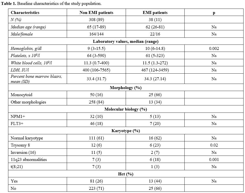

Table 1 Table 1. Baseline characteristics of the study population.

In the subgroup of patients with EMI, 24/38 were diagnosed with extramedullary involvement at diagnosis (63%), 10/38 had an extramedullary relapse (26%), while 4/38 patients had extramedullary disease both at diagnosis and at relapse (11%). Sites of EMI were: skin (22 patients, 58%), CNS (6 patients, 16%), lymph nodes (1 patient, 3%), pleura (2 patients, 5%), spleen (1 patient, 3%), bone (1 patient, 3%), peritoneum (1 patient, 3%). Four patients had multiple localizations (11%) (skin, CNS, bones, pancreas, breast, pleural). (Figure 1) All patients with CNS involvement presented signs and symptoms related to localization represented by facial nerve paresis in 5 cases and diplopy in 1 case.

Figure 1 
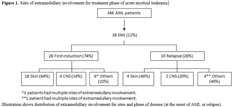
Figure 1. Sites of extramedullary involvement for treatment phase of acute myeloid leukemia.

Median age was 65 years (18-89) for AML patients without and 62 (26-81) for patients with EMI (p-value = ns). The median value of hemoglobin for AML patients without EMI was 9 g/dl (range 3-15.5), while for EMI patients resulted 10 g/dl (range 6-14.8) [p- value 0.002]; the median WBC count was 11x109/L  for AML patients without EMI (0.7-400) and 11.5x10⁹/L (1.3-272) for EMI ones (p-value: ns). The mean percent bone marrow blasts were 33% for patients with AML (+/- 31%) and 34% (+/- 27%) for EMI patients (p-value: ns) There were no differences in the average percentage of bone marrow blasts among EMI-negative and EMI-positive patients.
The morphology was monocytoid in 16% of AML patients without EMI (50 patients), and in 66% of EMI patients (25 patients) (p-value = 0.0001).
At flow cytometry analysis, blasts of patients diagnosed with EMI often lacked the expression of CD117: 15/38 patients with EMI (43%) were CD117 negative, versus 37/308 (19%) AML patients without EMI (p-value = 0.0035). 24 AML patients without EMI (12%) vs 9 EMI patients (26%) were CD56 positive (p = 0.061); 28 AML patients (14%) vs 10 (29%) EMI patient were both CD56 negative and CD117 negative (p = 0.046); 9 AML patients (5%) vs 5 (14%) EMI patients were at the same time CD56 positive and CD117 negative (p = 0.42) (Table 2).


Table 2 
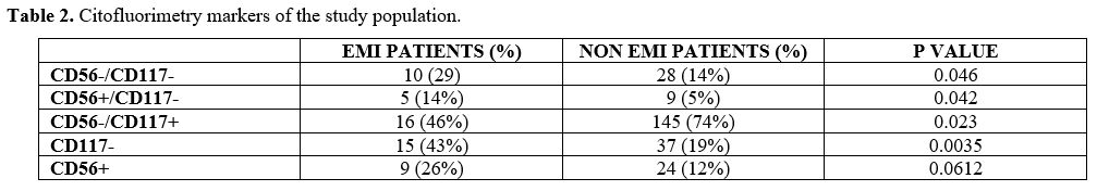
Table 2. Citofluorimetry markers of the study population.

The most frequent cytogenetic anomaly in EMI patients was trisomy 8 (23% of EMI patients vs 6% AML; p = 0.02), while t(8;21) and inv(16) were not significantly associated with EMI in our series (5% vs 7% for inv16; 3% vs 3% for t(8;21)).
NPM1 mutation was not more frequent in AML patients without EMI than EMI ones (10% vs 13%); similarly, 46/308 (18%) AML patients had FLT3 mutation vs 7/38 (20%) EMI patients (p-value: 0.78).
Thirteen patients among 346 AML patients (3.8%) had MLL rearrangements; 6/13 (46%) has been diagnosed with EMI (p = 0.001): 4 had leukemia cutis, one had a cutaneous and meningeal disease, and one had CNS localization only.
Further analysis regarding treatment, OS, and DFS was performed only on the 28 patients who experienced EMI at the onset of their disease. One EMI elderly patient, judged unfit for chemotherapy or hypomethylating agents due to age and comorbidities, received best supportive care only and was consequently excluded from OS analysis. Among the other EMI patients, 21 (55%) were treated with conventional chemotherapy, 5 with hypomethylating agents (13%), and 1 with low doses of cytarabine (3%).
Only eight patients (28.5%) were considered eligible, by age and absence of significant comorbidities, for a consolidation therapy with allogeneic stem cell transplantation (allo-HSCT) from a matched sibling (6 cases) or unrelated donor (2 cases): 5 patients received a myeloablative conditioning regimen, and three patients received a reduced-intensity conditioning regimen (RIC). Among the 27 patients with EMI included in the analysis, only 6 (22%) achieved CR versus an overall response rate of 123/163 AML patients without EMI (75 %) receiving similar therapeutic approaches (p-value < 0.0085).
No differences in DFS between EMI patients (7.4 months) and non-EMI AML patients (14.7 months) (p-value = 0.45) were observed.
The median OS of the 27 EMI patients was 11.6 months (2-79) (Figure 2).


Figure 2 
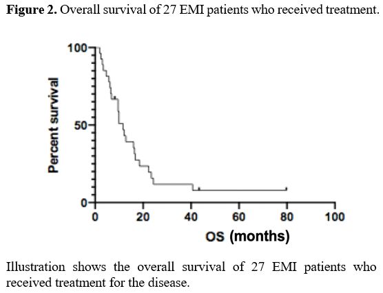
Figure 2. Overall survival of 27 EMI patients who received treatment.

Focusing on OS of AML patients treated with standard chemotherapy, not significant differences emerged between OS of 21 EMI patients (12 months) versus OS of 163 AML patients without EMI treated with a similar approach (15.8 months) (p-value = 0.09).
On the other hand, the OS of EMI patients who undergone allo-HSCT (16.7 months) resulted significantly different from OS of EMI patients who did not receive allo-HSCT (8.2 months) (p-value was 0.02). (Figure 3)
Both univariate and multivariate analyses showed that the achievement of CR and allo-HSCT were the main prognostic factors for survival in the EMI population (p < 0.0001, IC 0.39-0.63; p < 0.0001, IC 0.25-0.48 respectively).


Figure 3 
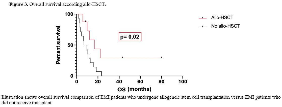
Figure 3. Overall survival according allo-HSCT.


Discussion

EMI is reported in 2.5%-9.1% of patients with AML and occurs concomitantly, following, or, rarely, antedating the onset of systemic bone marrow leukemia.[2] The most common locations include soft tissue, bone, gastrointestinal tract, and lymph nodes; the affection of the central nervous system is rare, with a reported frequency of 1.5%, which can have a crucial impact on the clinical course of the disease.[10]
The overall incidence of EMI in our study is 11% but drops to 7% if considered at AML diagnosis. The appearance of EMI in the course of AML is a complex phenomenon, and it is associated with a series of clinical and laboratory characteristics, such as high levels of lactate-dehydrogenases (LDH) and leukocytosis.[11] In our series, neither LDH levels nor leukocyte count was significantly associated with EMI.
It is also described a higher incidence of EMI in patients with either a myelomonocytic or a monocitoyd differentiation of leukemia cells;[12] this data was confirmed by our study also, where the rate of monocytoid differentiation of AML was significantly higher in EMI patients than in patients without EMI.
As far as the disease's molecular biology is concerned, EMI has been described in association with mutations of NPM1, FLT3, and MLL rearrangements.[13,14,15] We confirm that in our series, MLL mutations were frequently observed among EMI patients. Rearrangements of 11q23 are found in 11% of adult patients with AML and 20% of patients with AML and CNS involvement.[11] Martinez-Climent et al. showed the relationship between MLL anomalies and EMI: in their study on a pediatric population of 36 patients with AML, 11q23 rearrangements were significantly associated with leukocytosis, skin lesions, CNS localization of the disease, and a generally worse prognosis.[16] Interestingly in our study, 67% of patients with MLL rearrangement and EMI had skin involvement, associated with CNS localization in 16% of cases and high LDH levels; unlike the study previously reported, in our series, MLL rearrangement did not impact on prognosis.
Multiple chromosomal anomalies are reported as associated with extramedullary leukemia. Some common abnormalities are t(8,21) (q22;q22), inv(16), 11q23, t(9;11), t(8;17), t(8;16), t(8;17), t(1;11), trisomy of chromosomes 4, 8, 11, monosomy 7, and deletions of chromosomes 5q, 16q, and 20q. Complex karyotype occurs in about 17%-39% of patients.[17] In our series, no difference for complex karyotype was observed between EMI and non-EMI patients; differently trisomy of chromosome 8, which was the most frequent cytogenetic finding, was significantly more frequent in EMI patients.
Factors significantly associated with EMI in our study (i.e., trisomy 8, MLL rearrangements, and monocytoid differentiation  of  AML) confirm the results of  a  study by Laursen et al.,[18] which had described a correlation among trisomy 8, monocitoyd morphology, and MLL rearrangements in this kind of patients. 
The prognostic impact of cytogenetic alterations in the presence of EMI has not been delineated; Byrd et al. showed that extramedullary leukemia adversely affected hematologic complete remission rate and overall survival in patients with t(8;21)(q22;q22).[19]  In our series among eight patients with t(8;21), only one patient had EMI, without any significant CR and OS differences than the other patients without EMI. 
The mechanism for EMI is not fully understood, but homing to extramedullary tissues may be altered due to the blast expression of different adhesion molecules. CD56 has been described as a cytofluorimetry risk factor for EMI, and it is also common in patients with t(8;21) and monocytic or myelomonocytic morphology. Neural cell adhesion molecule is also highly expressed in breast, testicular, ovarian, and gut tissue, which could be EMI sites. Moreover, according to Bask et al., it has been described that the deregulation of CBF transcription factors in patients with inv(16) may play an important part in the pathogenesis of extramedullary involvement.[2] The lack of CD117 (c-kit) on the blast's surface has also been described as a possible enhancement of leukemic cell migrations in other sites than the bone marrow.[20] In our study, the cytofluorimetric analysis of bone marrow samples showed that 43% of EMI patients were negative for CD117.
Historically, extramedullary involvement has always been considered an adverse prognostic factor for adult patients with AML.[21] In our series, the overall survival was not significantly different when comparing EMI patients to the rest of the population enrolled in this study. On the other hand, EMI had a negative impact on disease-free survival (DFS), which was significantly shorter in EMI patients.
According to more recent data, the impact of EMI depends on the site involved and the other biological and cytogenetic characteristics of the disease.[3]
Among factors that negatively influence EMI prognosis is above all the kind of extramedullary location: patients with CNS involvement with the same cytogenetic and molecular abnormalities compared to patients with AML without EMI reach less frequently CR and have a 5-year lower survival.[7,11] Our data shows that the site of EMI had no significant impact on OS and DFS; in particular, although CNS involvement is generally considered an adverse prognostic factor,[11] in our series OS of patients with neuromeningeal disease did not significantly differ from the OS of other EMI patients.
Our data on OS in EMI patients are in line with those of Ganzel et al., who demonstrated that neither the presence of EMI nor the number of specific sites of EMI  influenced prognosis individually in a multivariable analysis adjusted for known prognostic factors such as cytogenetic risk and WBC count, in a population of 3522 adult patients with AML.[22]
There is no consensus on the treatment of AML patients with EMI because of the disease's rarity and the lack of randomized controlled studies.[17] In most single-institution series previously published, AML-like induction therapy, followed by consolidation with either chemotherapy or allo-HSCT, is the current standard of care in fit patients.[23]
Kaur et al.,[23] in a retrospective review of 23 patients treated for EMI, reported that the overall survival was significantly improved for patients who achieved a complete response to induction chemotherapy. This data was also confirmed in our study, where complete remission achievement after induction chemotherapy emerged as one of the most significant factors in multivariate analysis.
The possibility to perform allo‐HCT in our series showed to impact outcomes of patients positively.
Previous retrospective studies have demonstrated superior outcomes using allogeneic or autologous stem cell transplant in EMI.[24] Bourlon et al.[1] investigated the impact of EMI at diagnosis on the outcome of 39 patients transplanted for AML in first complete remission; this study concluded that EMI at the diagnosis of AML did not seem to influence outcomes following allo-HSCT performed in first CR.
Interestingly Goyal et al. found that the presence of extramedullary disease at any time before allo-HSCT did not adversely affect the outcomes, in terms of OS, leukemia-free survival, treatment-related mortality, or risk of relapse, in a large series of 814 EMI patients when compared with a cohort of AML patients without EMI.
The relatively small cohort size of transplanted patients in our study did not allow conclusions about better conditioning regimens in EMI patients.

Conclusions

From our study results, it has emerged that EMI incidence is not a rare event and affects a significant percentage of adult individuals with AML. In our case history, 11% of patients developed an extracellular localization of disease at onset and/or recurrence.
Some features, as trisomy 8, CD117 negative in immunophenotype, and monocytoid differentiation and MLL rearrangement, could be risk factors for EMI in AML patients.
The presence of an EMI does not in itself determine a reduced survival.
The achievement of a complete response after treatment and the possibility of performing an allogeneic transplant is confirmed to be the main characteristics that allow us to identify patients with more prolonged survival.


References   

  1. Bourlon C, Lipton JH, Deotare U, Gupta V, Kim DD, Kuruvilla J, Viswabandya A, Thyagu S, Messner HA, Michelis FV. Extramedullary disease at diagnosis of AML does not influence outcome of patients undergoing allogeneic hematopoietic cell transplant in CR1. Eur J Haematol. 2017;99(3):234-239. https://doi.org/10.1111/ejh.12909 PMid:28556258
  2. Bakst RL, Tallman MS, Douer D, Yahalom J. How I treat extramedullary acute myeloid leukemia. Blood. 2011;118(14):3785-3793. https://doi.org/10.1182/blood-2011-04-347229 PMid:21795742
  3. Zhang XH, Zhang R, Li Y. Granulocytic sarcoma of abdomen in acute myeloid leukemia patient with inv(16) and t(6;17) abnormal chromosome: Case report and review of literature. Leukemia Research. 2010;34(7): 958-961. https://doi.org/10.1016/j.leukres.2010.01.009 PMid:20116851
  4. Lazzarotto D, Candoni A, Filì C, Forghieri F, Pagano L, Busca A, Spinosa G, Zannier ME, Simeone E, Isola M, Borlengi E, Melillo L, Mosna F, Lessi F, Fanin R. Clinical outcome of myeloid sarcoma in adult patients and effect of allogeneic stem cell transplantation. Results from a multicenter survey. Leuk Res. 2017; 53:74-81. https://doi.org/10.1016/j.leukres.2016.12.003 PMid:28056398
  5. Slomowitz SJ, Shami PJ. Management of extramedullary leukemia as a presentation of acute myeloid leukemia. J Natl Compr Canc Netw. 2012;10(9):1165-1169. https://doi.org/10.6004/jnccn.2012.0120 PMid:22956813
  6. Agis H, Weltermann A, Fonatsch C. A comparative study on demographic, hematological, and cytogenetic findings and prognosis in acute myeloid leukemia with and without leukemia cutis. Ann Hematol 2002; 81:90-95. https://doi.org/10.1007/s00277-001-0412-9 PMid:11907789
  7. Del Principe MI, Buccisano F, Soddu S, Maurillo L, Cefalo M, Piciocchi A, Consalvo MI, Paterno G, Sarlo C, De Bellis E, Zizzari A, De Angelis G, Fraboni D, Divona M, Voso MT, Sconocchia G, Del Poeta G, Lo Coco F, Arcese W, Amadori S, Venditti A. Involvement of central nervous system in adult patients with acute myeloid leukemia: Incidence and impact on outcome. Semin Hematol. 2018;55(4):209-214. https://doi.org/10.1053/j.seminhematol.2018.02.006 PMid:30502849
  8. Döhner H, Estey E, Grimwade D, Amadori S, Frederick RA, Büchner T, Dombret H, Ebert BL, Fenaux P, Larson RA, Levine RL, Lo-Coco F, Naoe T, Niederwieser D, Ossenkoppele GJ, Sanz M, Sierra J, Tallman MS, Tien HF, Wei AH, Löwenberg B, Bloomfield CD. Diagnosis and management of AML in adults: 2017 ELN recommendations from an international expert panel. Blood. 2017;129(4):424-447. https://doi.org/10.1182/blood-2016-08-733196 PMid:27895058 PMCid:PMC5291965
  9. Cheson BD, Bennett JM, Kopecky KJ, Büchner T, Willman CL, Estey EH, Schiffer CA, Doehner H, Tallman MS, Lister TA, Lo-Coco F, Willemze R, Biondi A, Hiddemann W, Larson RA, Löwenberg B, Sanz MA, Head DR, Ohno R, Bloomfield CD. Revised recommendations of the International Working Group for Diagnosis, Standardization of Response Criteria, Treatment Outcomes, and Reporting Standards for Therapeutic Trials in Acute Myeloid Leukemia [published correction appears in J Clin Oncol. 2004 Feb 1;22(3):576 J Clin Oncol. 2003;21(24):4642-4649. https://doi.org/10.1200/JCO.2003.04.036 PMid:14673054
  10. Meyer, H, Pönisch W, Schmidt SA, Wienbeck S, Braulke F, Schramm D, Surov A. Clinical and imaging features of myeloid sarcoma: a German multicenter study. BMC Cancer 19, 1150 (2019). https://doi.org/10.1186/s12885-019-6357-y PMid:31775680 PMCid:PMC6882227
  11. Alakel N, Stölzel F, Mohr B, Kramer M, Oelschlägel U, Rölling C, Bornhäuser M, Ehninger G, Schaich M. Symptomatic central nervous system involvement in adult patients with acute myeloid leukemia. Cancer Manag Res. 2017; 9:97-102. Published 2017 Mar 29. https://doi.org/10.2147/CMAR.S125259 PMid:28435324 PMCid:PMC5386598
  12. Holmes R, Keating MJ, Cork A, Broach Y, Trujillo J, Dalton Jr WT, McCredie KB, Freireich EJ. A unique pattern of central nervous system leukemia in acute myelomonocytic leukemia associated with inv(16)(p13q22). Blood. 1985;65(5):1071-1078. https://doi.org/10.1182/blood.V65.5.1071.1071 PMid:3857943
  13. Ansari-Lari MA, Yang CF, Tinawi-Aljundi R, Cooper L, Long P, Allan RH, Borowitz MJ, Berg KD, Murphy KM. FLT3 mutations in myeloid sarcoma. Br J Haematol. 2004 Sep;126(6):785-91. https://doi.org/10.1111/j.1365-2141.2004.05124.x PMid:15352981
  14. Falini B, Lenze D, Hasserjian R,Coupland S, Jaehne D, Soupir C, Liso A, Martelli MP, Bolli N, Bacci F, Pettirossi V, Santucci A, Martelli MF, Pileri S, Stein H. Cytoplasmic mutated nucleophosmin (NPM) defines the molecular status of a significant fraction of myeloid sarcomas. Leukemia. 2007;21(7):1566-1570. https://doi.org/10.1038/sj.leu.2404699 PMid:17443224
  15. Ohanian M, Faderl S, Ravandi F, Pemmarajiu N, Garcia-Manero G, Cortes J, Estrov Z. Is acute myeloid leukemia a liquid tumor? Int J Cancer. 2013;133(3):534-543. https://doi.org/10.1002/ijc.28012 PMid:23280377 PMCid:PMC3904286
  16. Martinez-Climent JA, Espinosa R, Thirman MJ, Le Beau MM, Rowley JD. AML with 11q23/MLL abnormalities as defined by the WHO classification: incidence, partner chromosomes, FAB subtype, age distribution, and prognostic impact in an unselected series of 1897 cytogenetically analyzed AML cases. J Pediatr Hematol Oncol. 1995 Nov;17(4):277-83.
  17. Shanin O, Ravandi F. Myeloid sarcoma. Current opinion in Hematologu. 2020; 27(2); 88-94. https://doi.org/10.1097/MOH.0000000000000571 PMid:31904666
  18. Laursen L AC, Sandahl D J, Kjeldsen E, Abrahamsson J, Asdahl P, Ha SY, Heldrup J, Jahnukainen K, Jònsson OG, Lausen B, Palle J, Zeller B, Forestier E, Hasle H. Trisomy 8 in Pediatric Acute Myeloid Leukemia: A NOPHO AML Study. 2016. GENES, CHROMOSOMES and CANCER 55(9): 719-26. https://doi.org/10.1002/gcc.22373 PMid:27153159
  19. Byrd JC, Weiss RB, Arthur DC, Lawrence D, Baer MR, Davey F, Trikha ES, Carroll AJ, Tantravahi R, Qumsiyeh M, Patil SR, Moore JO, Mayer RJ, Schiffer CA, Bloomfield CD. Extramedullary leukemia adversely affects hematologic complete remission rate and overall survival in patients with t(8;21)(q22;q22): results from Cancer and Leukemia Group B 8461. J Clin Oncol. 1997 Feb;15(2):466-75. https://doi.org/10.1200/JCO.1997.15.2.466 PMid:9053467
  20. Liesveld JL. Expression and function of adhesion receptors in acute myelogenous leukemia: parallels with normal erythroid and myeloid progenitors. Acra Haematol 1997; 97:53-62. https://doi.org/10.1159/000203659 PMid:8980610
  21. Goyal SD, Zhang M-J, Wang H-L, Akpek G, Copelan EA, Freytes C. Allogenic hematopoietic cell transplant for AML: no impact of pre-transplant extrammedullary disease on outcome. Bone Marrow Transplantation (2015) 1-6.
  22. Ganzel C, Manola J, Douer D, Rowe JM, Fernandez HF, Paietta EM, Litzow MR, Lee JW, Luger SM, Lazarus HM, Cripe LD, Wiernik PH, Tallman MS. Extramedullary Disease in Adult Acute Myeloid Leukemia Is Common but Lacks Independent Significance: Analysis of Patients in ECOG-ACRIN Cancer Research Group Trials, 1980-2008 [published correction appears in J Clin Oncol. 2017 Jan 10;35(2):263]. J Clin Oncol. 2016;34(29):3544-3553. https://doi.org/10.1200/JCO.2016.67.5892 PMid:27573652 PMCid:PMC5074349
  23. Kaur V, Swami A, Alapat D, Abdallah AO, Motwani P, Hutchins LF Jethava Y. Clinical characteristics, molecular profile and outcomes of myeloid sarcoma: a single institution experience over 13 years. Hematology, 2017. https://doi.org/10.1080/10245332.2017.1333275 PMid:28574302
  24. Chevallier P, Mohty M, Lioure B, Michel G, Contentin N, Deconinck E, Bordigoni P, Vernant JP, Hunault M, Vigouroux S, Blaise D, Tabrizi E, Buzyn A, Socie G, Michallet M, Volteau C, Harousseasu L. Allogeneic hematopoietic stem-cell transplantation for myeloid sarcoma: a retrospective study from the SFGM-TC. J Clin Oncol. 2008;26(30):4940-3. https://doi.org/10.1200/JCO.2007.15.6315 PMid:18606981

[TOP]