Phenotypic and Molecular Detection of Biofilm Formation in Methicillin-Resistant Staphylococcus Aureus Isolated from Different Clinical Source in Erbil City

Pishtiwan Ahmad Hamad.

Biology Department, College of Education, Salahaddin University-Erbil, Iraq

Correspondence to: Pishtiwan Ahmad Hamad. Biology Department, College of Education, Salahaddin University-Erbil, Iraq. Email: Pishtiwan.hamad@su.edu.krd

Published: March 1, 2023
Received: November 10, 2022
Accepted: February 16, 2023
Mediterr J Hematol Infect Dis 2023, 15(1): e2023016 DOI 10.4084/MJHID.2023.016

This is an Open Access article distributed under the terms of the Creative Commons Attribution License
(
https://creativecommons.org/licenses/by-nc/4.0), which permits unrestricted use, distribution, and reproduction in any medium, provided the original work is properly cited.

Abstract

Background: Staphylococcus aureus is an important causative pathogen. The production of biofilms is an important factor and makes these bacteria resistant to antimicrobial therapy.
Objectives:
The current study aimed to assess the prevalence of resistance to antibacterial agents and to evaluate the phenotypic and genotypic characterization of biofilm formation among S. aureus strains.
Methods:
This study included 50 isolates of Methicillin-Resistant S. aureus (MRSA) and Methicillin-Susceptible S. aureus (MSSA). S. aureus was identified by molecular and conventional methods, and Antimicrobial resistance was tested with a disc diffusion method. The biofilm formation was performed through the Microtiter plate method. Strains were subjected to PCR to determine the presence of nuc, mecA, icaA, icaB, icaC and icaD genes.
Results:
Of the 50 S. aureus isolates, 32(64%) and 18(36%) were MRSA and MSSA, respectively. A large number of MRSA and MSSA isolates showed resistance to Penicillin and Azithromycin, and a lower number of MRSA and MSSA isolates showed resistance to Amikacin Gentamicin. None of the isolates was resistant to Vancomycin. The MRSA strains had significantly higher resistance against antibiotics than MSSA strains (P = 0.0154). All isolates (MRSA and MSSA) were able to produce biofilm with levels ranging from strong (31.25 %), (16.6%) to moderate (53.12%), (50%) to weak (15.6 %), (33.3% %) respectively. The MRSA strains had a significantly higher biofilm formation ability than the MSSA strains (P = 0.0079). The biofilm-encoding genes were detected among isolates with different frequencies. The majority of S. aureus isolates, 42 (84%), were positive for the icaA. The prevalence rates of the icaB, icaC and icaD genes were found to be 37 (74%), 40 (80%) and 41 (82%), respectively.
Conclusions:
The prevalence of biofilm encoding genes associated with multidrug resistance in S. aureus strains is high. Therefore, identifying epidemiology, molecular characteristics, and biofilm management of S. aureus infection would be helpful.


Introduction

The most prevalent agents of the hospital- and community-acquired infections are Staphylococcus aureus (S. aureus). Furthermore, one of the main human pathogens is methicillin-resistant  (MRSA). It is a bacterial pathogen that causes many illnesses, including skin infections, dangerous invasive infections like pneumonia, soft tissue infections, bones, heart valves, and even deadly human septicemia. It also quickly develops antibiotic resistance.[1,2] In recent years, S. aureus infections have become more deadly due to the increasing incidence of antimicrobial resistance in S. aureus due to extensive antibiotic use.[3] The risk of death and the treatment cost were higher in the infections caused by antibiotic-resistant strains than by susceptible strains.[4] Antimicrobial resistance in methicillin-resistant strains of S. aureus (MRSA) is linked with the acquisition of a mobile genetic element named the staphylococcal cassette chromosome mec, which carries the mecA gene, encoding the low-affinity penicillin-binding protein 2a and confers resistance to the β-lactam antibiotics.[5] Although MRSA are resistant to β-lactam antibiotics, many MRSA isolates are multidrug-resistant (MDR) because they exhibit resistance to other antimicrobial agents, such as macrolides, tetracycline, aminoglycosides, chloramphenicol, and fluoroquinolones, which are frequently used in the treatment of infections caused on by these microorganisms.[6]
In addition to the bacteria's resistance to antibiotics, its ability to develop biofilm, a dynamic structurally complex multilayered cellular matrix, is another significant complicated factor for a better understanding of the molecular pathogenesis of S. aureus. As a result, new preventative and therapeutic approaches may be developed. Biofilm production, which is required for the survival and persistence of MRSA in its hosts, is considered to be a significant virulence factor, as well as one of many, including extracellular toxins and surface features that is effective in the induction and maintenance of infection in the host.[7] Initial attachment, biofilm maturation, and dispersal are the three main processes that can be classified as stages in the evolution of biofilms, according to several definitions. An individual planktonic cell will associate reversibly with a surface during initial attachment, and if it does not disassociate, it will bond irrevocably to the surface. Attachment is facilitated through surface proteins, referred to as microbial surface components recognizing adhesive matrix molecules (MSCRAMMs).[8] During infection, these proteins play major roles in attachment to host factors such as fibrinogen, fibronectin, and collagen. Biofilm maturation occurs through cell division and the production of the extracellular polymeric matrix. The composition of the biofilm matrix varies between strains but generally can contain host factors, polysaccharides, proteins, and extracellular DNA (eDNA).[9,10] Following biofilm accumulation, cells within the biofilm can reactivate to a planktonic state through dispersal.[11]
Production of biofilms is crucial during infection because it protects the bacteria against various human defense mechanisms and shields them from antimicrobial agents.[12] The ability to form biofilm is a trait associated with bacterial virulence and many chronic bacterial infections.[13] Several genes are involved in the production and maintenance of biofilms by staphylococci, of which the most widely studied are the icaA and icaD (intercellular adhesion A and B) genes responsible for the production of polysaccharide intercellular adhesion (PIA) that includes N-acetyl glucosamine as a primary component of the exopolysaccharide matrix surrounding the bacterial cells within the biofilm.[14] The aim of this study was to determine antimicrobial resistance profiles and detection of biofilm formation of MRSA and MSSA isolates from different clinical sources from patients in hospitals of Erbil city-Iraq.

Materials and Methods

Bacterial Strains. A total of 50 S. aureus strains from a clinical source, such as swabs from urine (25), a wound (10), and a catheter (10) throat (5), were used in this work. The strains were obtained from hospitals in Erbil City, Erbil, Iraq. The isolates were identified using traditional microbiological techniques. Besides, the VITEK 2 compact system was utilized to reidentify them (BioMerieux, France). Finally, the isolates' identities were verified using polymerase chain reaction (PCR) (Alpha PCRmax, UK) by detecting nuc genes. Table 1 contains the gene sequences and PCR setup.

Table 1
Table 1. Sequences of oligonucleotide primers used for PCR amplification of biofilm-associated genes with nuc and mecA genes used in this study.

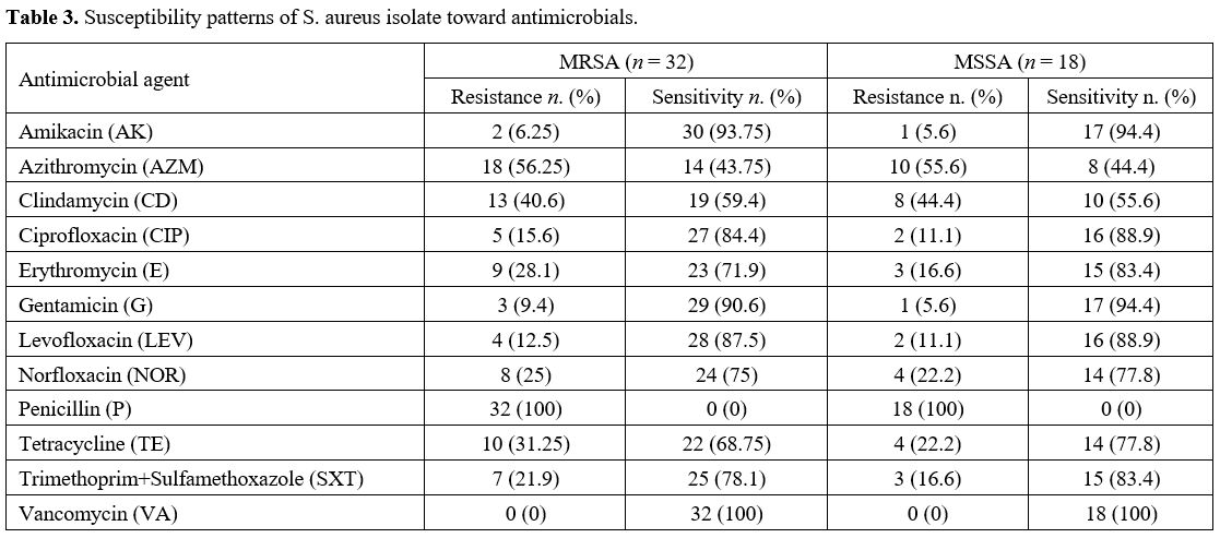
Antibiotic Susceptibility Testing. The disc diffusion technique was used to test all staphylococcal isolates for methicillin resistance. PCR verified methicillin resistance to detect the mecA gene.[16] The isolates' susceptibility was evaluated using the disc diffusion technique[20] using the following antibiotic discs (Bioanalyse, Turkey): Amikacin AK 30 μg, Azithromycin AZM 15 μg, Clindamycin CD 2 μg, Ciprofloxacin CIP 5 μg, Erythromycin E 15 μg, Gentamicin G 10 μg, Levofloxacin LEV 5 μg, Norfloxacin NOR 10 μg, Penicillin P 10 U, Tetracycline TE 30 μg, Trimethoprim+Sulfamethoxazole SXT 1.25+23.75 μg, and Vancomycin VA 30 μg. S. aureus ATCC 25923 was used as the control strain.
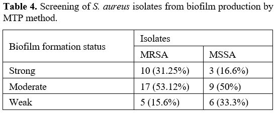
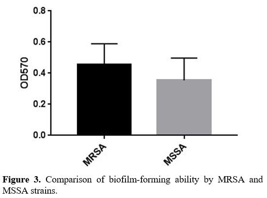
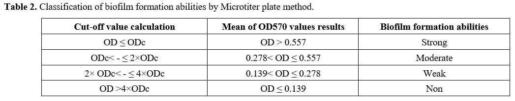
Biofilm Formation Assay. Biofilm generation was quantified using a Microtiter plate (MTP) approach described by Yousefi M. et al.[21] In brief, bacterial isolates were cultured in trypticase soy broth (TSB) (Merck, Germany) with 0.5 percent glucose and incubated at 37ºC overnight. Cultures with 0.5 percent glucose with 1:40 in fresh TSB were diluted (Sigma, USA). Two hundred μL of the diluted solution was put into Microtiter plate wells and incubated for 48 hours at 37ºC. Only 200 μL of TSB-0.5% glucose was present in the negative control wells, and there was no bacterial suspension. Wells were carefully cleaned three times with phosphate buffer saline (PBS) (pH 7.2), fixed for 20 minutes with methanol, dried at room temperature, and stained for 10 minutes with crystal violet 0.1 percent. Then 1 mL of 95% ethanol was added to each well to dissolve the dye bound to the adherent cells. Finally, using an ELISA reader (BioTek ELx800, USA), optical density (OD) at 570 nm (A570) was determined for each well. The average OD of negative control + 3 standard deviation (SD) of negative control was calculated for the optical density cut–off (ODc). Based on the absorbance of crystal violet stain linked to the adhered cells, biofilms formed by various strains have been analyzed and categorized (Table 2).


Table 2
Table 2. Classification of biofilm formation abilities by Microtiter plate method.

DNA Isolation. DNA extraction from a genome following the manufacturer's instructions, genomic DNA was isolated from pure cultures using the Presto™ Mini gDNA Bacteria Kit (Geneaid, Taiwan); the extract was then eluted with a 100 μL elution buffer. Extracts were kept at -20°C until PCR was performed. A DNA template was used for PCR using 2 μL of the total extracted material from each test sample.
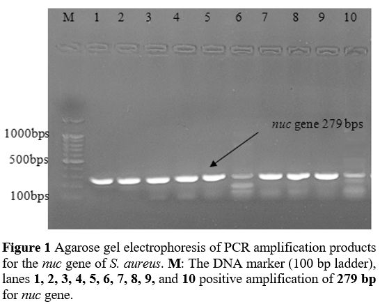
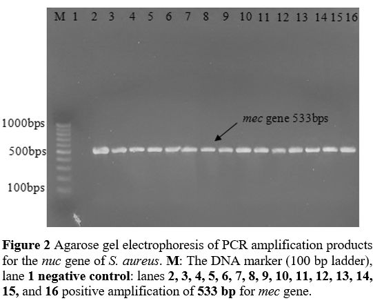
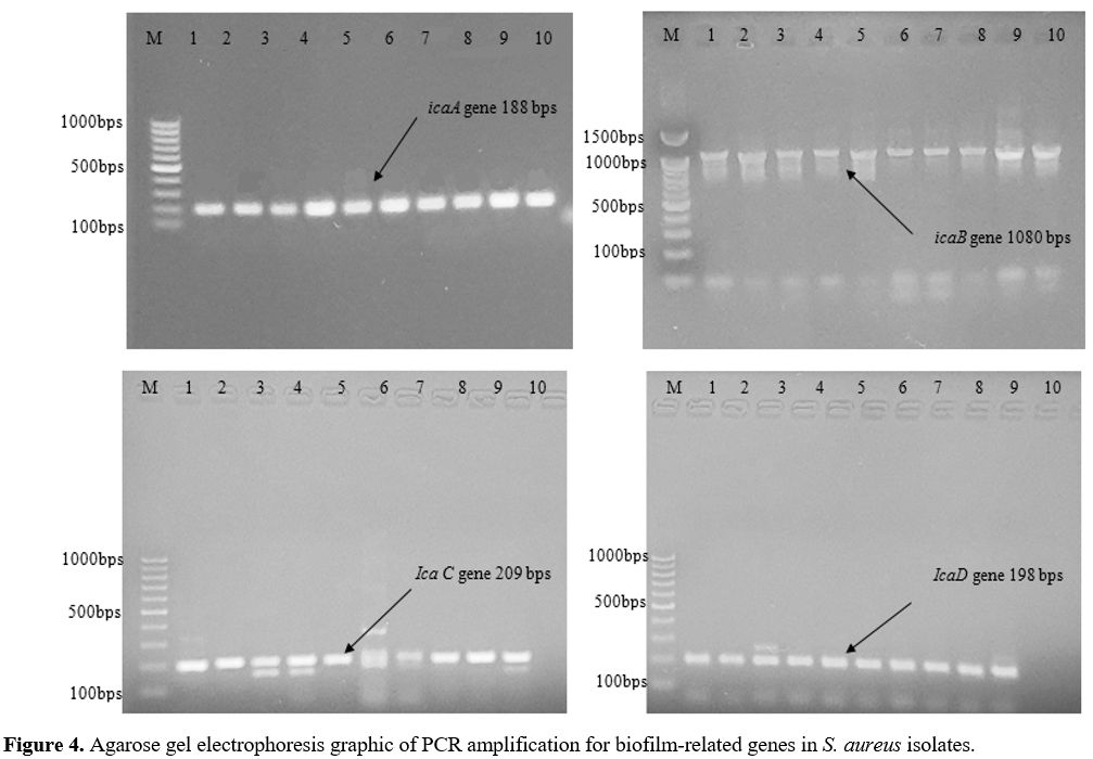
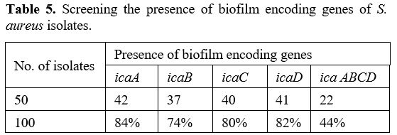
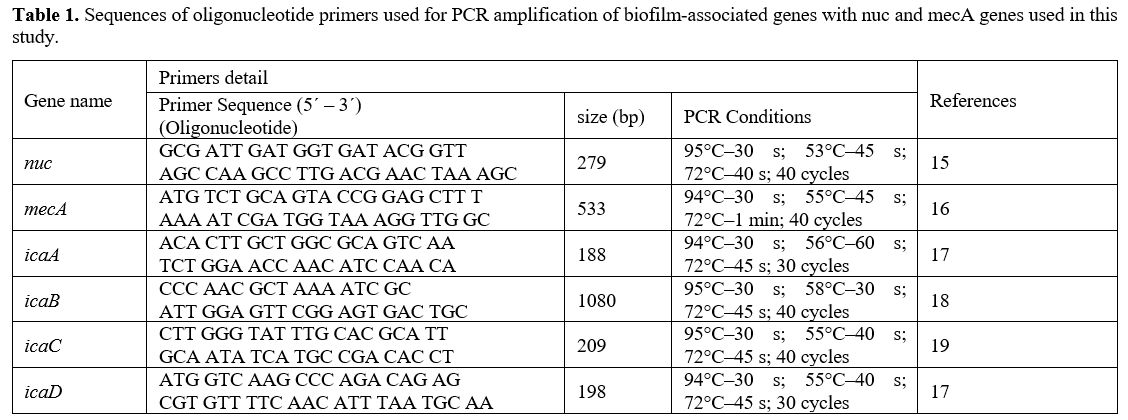
Primers and PCR Conditions. The primers designed by BioGene (South Korea) specifically for the nuc, mecA, icaA, icaB, icaC, and icaD are given in Table 2. The monoplex PCR was performed in a 25-𝜇L volume for each gene. All PCR reactions were conducted by employing 2 μl DNA template (density of 10 ng/μl), a Master Mix comprising of 3 mM MgCl2, 0.2% Tween® 20, 20 mM Tris-HCl pH 8.5, (NH4)2S04, 0.4 mM of each dNTP, 0.4 μM of each primer, and 0.2 units/μl Ampliqon Taq DNA polymerase. The PCR amplification conditions were established as shown in Table 1.
Statistical Analysis. The T-test was used to examine group differences at the 0.05 level of significance. Utilizing the statistical analysis program GraphPad Prism 7.


Results

Isolation and characterization of S. aureus. 50 S. aureus isolates from patients at hospitals in Erbil City, Iraq, were used in this study and were identified using standard microbiological techniques. The VITEK II Compact System was used, and it reidentified all strains of S. aureus to support the identification of S. aureus isolates made by a standard approach. All isolates of S. aureus were further tested for the presence of the nuc genes to verify their identities; the presence of nuc genes in all of the strains proved that they were all S. aureus (Figure 1). Disk diffusion assay was used to test S. aureus isolates for methicillin resistance and separate them into MRSA (32 strains) and MSSA (18 strains). The mecA gene was detected by PCR to verify Methicillin resistance (Figure 2).

Figure 1 Figure 1. Agarose gel electrophoresis of PCR amplification products for the nuc gene of S. aureus. M: The DNA marker (100 bp ladder), lanes 1, 2, 3, 4, 5, 6, 7, 8, 9 and 10 positive amplification of 279 bp for nuc gene.

Figure 2 Figure 2. Agarose gel electrophoresis of PCR amplification products for the nuc gene of S. aureus. M: The DNA marker (100 bp ladder), lane 1 negative control: lanes 2, 3, 4, 5, 6, 7, 8, 9, 10, 11, 12, 13, 14, 15 and 16 positive amplification of 533 bp for mec gene.

Antibiotic susceptibility of S. aureus isolates.
- Antibiotic resistance pattern of the MRSA isolates. Of the total 50 S. aureus isolates, 32 (62%) were Methicillin-resistant 
S. aureus (MRSA). A large number of MRSA isolates showed resistance to Penicillin (100%), Azithromycin (56.25%), Clindamycin (40.6), and Tetracycline (31.25). A lesser number of MRSA isolates showed resistance to Amikacin (6.25%), Gentamicin (9.4%), Levofloxacin (12.5%), Ciprofloxacin (15.6%), trimethoprim with sulfamethoxazole (21.9%), Norfloxacin (25%), and Erythromycin (28.1%). Vancomycin resistance was not seen in any MRSA samples (Table 3). 

Table 3
Table 3. Susceptibility patterns of S. aureus isolate toward antimicrobials

The MRSA isolates were screened for resistance to 9 different antimicrobial drug groups, and resistance to at least 3 groups revealed that the isolates were MDRs (multidrug-resistant). Out of 32 MRSA isolates, 19 (59.3%) were MDR (resistant to three or more antibiotics), 3 (9.3%) were resistant to only two antibiotics, and 10 (31.2%) were resistant to only one antibiotic. The most prevalent resistance pattern among MDR MRSA isolates, in addition to resistance to -lactam antibiotics, was resistance to azithromycin, clindamycin, and tetracycline.
- Antibiotic resistance pattern of the MSSA isolates. Eighteen isolates (36%) of the 50 Staphylococcus isolates were Methicillin-Susceptible 
S. aureus (MSSA). The rate of resistance of the 18 MSSA isolates to the antibiotics shows various sensitivity patterns (Table 4). The greatest observed resistance rate was against Penicillin (100%), Azithromycin (55.6%), and Clindamycin (44.4). Also, the lowest resistant percentage of MSSA isolates recorded was against Amikacin and Gentamicin (5.6), then Levofloxacin and Ciprofloxacin (11.1%), trimethoprim with sulfamethoxazole and Erythromycin (16.6%), Norfloxacin and Tetracycline (22.2) respectively. None of the MSSA isolates was resistant to Vancomycin (Table 2). The multidrug resistance pattern of the MSSA demonstrated that eleven (61.1%) of the MSSA were multidrug-resistant, two (11.1%) were resistant to only two antibiotics, and five (27.7%) were resistant to only one antibiotic (Table 3).

Table 4
Table 4. Screening of S. aureus isolates from biofilm production by MTP method.

A statistical test comparing the antibiotic resistance pattern between MRSA and MSSA isolates showed a significant difference between MRSA and MSSA isolates, and MRSA strains had significantly higher resistance against antibiotics than MSSA strains  (P = 0.0154).
Biofilm Formation Assay
. The MTP was used to assess 
S. aureus strains for their ability to produce biofilms. In this study, after crystal violet staining, OD570 mean microplate readings ranged from 0.186 to 0.613. The negative control mean was 0.054. Therefore, an ODc570 of biofilm production was defined as 0.139. The strains were classified into four categories: non–biofilm producer (−), OD570≤0.139; weak biofilm producer (+), 0.139<OD570≤0.278; moderate biofilm producer (++), 0.278<OD570≤0.557; strong biofilm producer (+++), 0.557≤OD570. S. aureus strains that were included in this work were divided into MRSA (32 strains) and MSSA (18 strains), and the information reveals that all S. aureus isolates tested positive for biofilms.
The majority of the 32 MRSA strains examined (n=17) (53.12%) produced moderate biofilms. The strong biofilms were produced by (n=10) 31.25 % of the MRSA strains, while (n=5) 15.6 % of strains were weak biofilm producers. A large group of the 18 MSSA strains examined (n=9) (50%) also formed moderate biofilms, while (n=3) 16.6% and (n=6) 33.3% of MSSA strains were strong and weak producers, respectively (Table 4).
Comparison of biofilm biomass (absorbance at 570 nm) Using a statistical analysis, it was determined that MRSA isolates were significantly more capable of forming biofilms than MSSA isolates (P = 0.0079) (Figure 3).


Figure 3
Figure 3  Comparison of biofilm-forming ability by MRSA and MSSA strains.

Detection of biofilm formation genes. The existence of the ica operon in the tested strains was verified by amplification of specific segments for icaA (188 bp), icaB (1080 bp), icaC (209 bp), and icaD (198 bp) Figure 4 and the relationship between the formation of biofilm and the four genes associated with biofilm was evaluated. These genes' distribution in S. aureus isolates is shown in Table 5. All four genes were found among isolates with different occurrences. As observed, a large percentage of S. aureus isolates, 42 (84%), were found to be icaA positive. On the other hand, the prevalence rates of the icaB, icaC, and icaD genes were unswervingly found to be 37 (74%), 40 (80%) and 41 (82%), respectively.

Figure 4 Figure 4. Agarose gel electrophoresis graphic of PCR amplification for biofilm-related genes in S. aureus isolates.

Table 5 Table 5. Screening the presence of biofilm encoding genes of S. aureus isolates.

Twenty-two 44% of isolates that produce biofilm were revealed to have all four genes. There are great relationships between strong biofilm formation in isolates and the presence of all four biofilm encoding genes. Also, a significant relationship was found between the presence of icaA (p= 0.0036), icaB (p= 0.0001), icaC (p= 0.001), and icaD (p= 0.0019) gene and phenotypic biofilm formation in S. aureus isolates. The occurrence of icaABCD genes was highly significantly associated with the phenotypic biofilm formation in S. aureus isolates (P = <0.0001).
 

Discussion

S. aureus is an opportunistic pathogen that has long been recognized as a frequent source of human infections. Despite being a natural component of human flora, S. aureus has the potential to cause a wide variety of illnesses, from relatively minor skin infections to catastrophic outcomes. Many of these illnesses can quickly become life-threatening if they are not properly controlled and treated.[22] The ability to form biofilm using some phenotypic and genotypic indicators is related to the spread of antibiotic resistance, especially resistance to methicillin in infected samples. It is one of the main concerns related to the ability of Staphylococcus aureus to threaten health. Considering the formation of multilayered units in the creation of biofilm structures, the expansion of biofilm samples can be considered a key and indicator step in the increase of infection and the expansion of antibiotic resistance. The objective of this study was to investigate the phenotypic and molecular diagnosis of biofilm formation in methicillin-resistant Staphylococcus aureus isolated from different clinical sources.
In this study, antibiotic susceptibility testing and mecA gene identification revealed that 32 strains were MRSA and 18 were MSSA. Kumurya et al. have reported similar validation procedures.[23] A large number of MRSA strains are sensitive to penicillin, azithromycin, clindamycin, tetracycline, sulfamethoxazole, gentamicin, levofloxacin, Norfloxacin, ciprofloxacin, Amikacin, as shown by the studies conducted by John Walter et al. and Stefanaki et al.[24,25]
In this study, the rate of resistance to erythromycin was 28.1%, which is not consistent with the study of J Szabó et al., and Romen Singh Naorem et al., which may be due to the different number of strains in the studies.[26,27] The insignificant resistance of MRSA strains to Vancomycin in this study is consistent with studies conducted by Vinay Kumar Moses et al. and Chaudhari CN et al.[22,28] The MSSA isolates were susceptible to most antibiotics tested. However, more resistance was observed to penicillin, azithromycin, clindamycin, and to some extent to clindamycin, tetracycline, tobramycin, Amikacin, gentamicin, ciprofloxacin, Norfloxacin, and levofloxacin. In contrast, in the case of MRSA, multiple drug resistance was common, and only a few antibiotics were active against these isolates. Also, the resistance of MSSA strains to Vancomycin was negligible and zero. These findings are consistent with the findings of Fateh Rahimi et al., Shilpa Arora et al., and H Saderi et al.[29-31] Comparing the two antibiotic resistance patterns of MRSA and MSSA isolates showed that MRSA strains have much higher antibiotic resistance than MSSA strains. This finding is consistent with the findings of Solmaz Dibah et al., and Ricciardi et al.[32,33]
S. aureus has been found to produce biofilms, which are thought to cause chronic or persistent infections because they act as a defense against the immune system and antibiotics.[6] This study used MTP to form the biofilm of Staphylococcus aureus strains. A study including 50 clinical isolates of Staphylococcus aureus reported that MTP is a more accurate and repeatable method for biofilm detection.[34] MRSA strains showed a higher percentage of biofilm formation (moderate and strong) than MSSA strains. Moreover, MSSA strains showed a higher percentage of forming weak biofilms. These findings are consistent with the study findings of Leshem et al. and Omidi et al.[35,36] In addition, the existence of icaABCD operon components is often correlated with biofilm generation, which is consistent with the findings of research by Shivaee et al. and Khasawneh et al.[37,38] In this study, as can be seen, most of the Staphylococcus aureus isolates (from low to high, icaC - icaB - icaD - icaA) were positive for biofilm-related genes. These findings are consistent with the findings of the studies of Mehdi Goudarzi et al. and Ali Haghi et al.[39,40] On the other hand, Azmi et al. pointed out that the icaA gene's low prevalence (16.5%)[41] suggests that alternative mechanisms may be used by these strains in addition to the ica-dependent system, which may not be the only mechanism involved in biofilm development.

Conclusions

The results of the present study have shown that Staphylococcus aureus isolates had high resistance to most of the investigated antibiotics. It seems that the biofilm in Staphylococcus aureus has a high phenotypic expression, and the high ability of this bacterium to form a biofilm can be particularly important in the emergence of infections and the creation of multiple antibiotic strains. Considering the importance of Staphylococcus aureus bacteria as a hospital pathogen and considering the increasing antibiotic resistance in clinical isolates in order to prevent the formation of biofilm and colonization of bacteria in hospital tools and environment, it is recommended to observe proper sterilization in the therapeutic tools related to the patient, to prevent the transmission of infection and also the formation of biofilm.

Acknowledgements

We want to thank all the patients who patiently helped in this research and also thank them for their support and help.
 

References   

  1. Pathare NA, Tejani S, Asogan H: Comparison of methicillin resistant Staphylococcus aureus in healthy community hospital visitors [CA-MRSA] and hospital staff [HA-MRSA]. Mediterr J Hematol Infect Dis 2015, 7:1-6. https://doi.org/10.4084/mjhid.2015.053 PMid:26543522 PMCid:PMC4621180
  2. Gordon RJ, Lowy FD: Pathogenesis of methicillin-resistant Staphylococcus aureus infection. Clinical infectious diseases 2008, 46:S350-S9. https://doi.org/10.1086/533591 PMid:18462090 PMCid:PMC2474459
  3. Neyra RC, Frisancho JA, Rinsky JL, Resnick C, Carroll KC, Rule AM, Ross T, You Y, Price LB, Silbergeld EK: Multidrug-resistant and methicillin-resistant Staphylococcus aureus (MRSA) in hog slaughter and processing plant workers and their community in North Carolina (USA). Environ Health Perspect 2014, 122:471-7. https://doi.org/10.1289/ehp.1306741 PMid:24508836 PMCid:PMC4014760
  4. Mulvey MR, Simor AE: Antimicrobial resistance in hospitals: how concerned should we be? Cmaj 2009, 180:408-15. https://doi.org/10.1503/cmaj.080239 PMid:19221354 PMCid:PMC2638041
  5. Kot B, Wierzchowska K, Piechota M, Grużewska A: Antimicrobial resistance patterns in methicillin-resistant Staphylococcus aureus from patients hospitalized during 2015-2017 in hospitals in Poland. Med Princ Pract 2020, 29:61-8. https://doi.org/10.1159/000501788 PMid:31256152 PMCid:PMC7024858
  6. Piechota M, Kot B, Frankowska-Maciejewska A, Grużewska A, Woźniak-Kosek A: Biofilm formation by methicillin-resistant and methicillin-sensitive Staphylococcus aureus strains from hospitalized patients in Poland. Biomed Res Int 2018, 2018:1-7. https://doi.org/10.1155/2018/4657396 PMid:30687745 PMCid:PMC6327255
  7. Lister JL, Horswill AR: Staphylococcus aureus biofilms: recent developments in biofilm dispersal. Front Cell Infect Microbiol 2014, 4:178-87. https://doi.org/10.3389/fcimb.2014.00178 PMid:25566513 PMCid:PMC4275032
  8. Foster TJ, Geoghegan JA, Ganesh VK, Höök M: Adhesion, invasion and evasion: the many functions of the surface proteins of Staphylococcus aureus. Nat Rev Microbiol 2014, 12:49-62. https://doi.org/10.1038/nrmicro3161 PMid:24336184 PMCid:PMC5708296
  9. Manoharan A, Premalatha K, Chatterjee S, Mathai D, Group SS: Correlation of TEM, SHV and CTX-M extended-spectrum beta lactamases among Enterobacteriaceae with their in vitro antimicrobial susceptibility. Indian J Med Microbiol 2011, 29:161-4. https://doi.org/10.4103/0255-0857.81799 PMid:21654112
  10. Cue DR, Lei MG, Lee C: Genetic regulation of the intercellular adhesion locus in staphylococci. Front Cell Infect Microbiol 2012, 2:38-51. https://doi.org/10.3389/fcimb.2012.00038 PMid:23061050 PMCid:PMC3459252
  11. Boles BR, Horswill AR: Staphylococcal biofilm disassembly. Trends Microbiol 2011, 19:449-55. https://doi.org/10.1016/j.tim.2011.06.004 PMid:21784640 PMCid:PMC3164736
  12. Scherr TD, Heim CE, Morrison JM, Kielian T: Hiding in plain sight: interplay between staphylococcal biofilms and host immunity. Front Immunol 2014, 5:37-44. https://doi.org/10.3389/fimmu.2014.00037 PMid:24550921 PMCid:PMC3913997
  13. Jamal A, David B: Biofilm formation by enterococci. J Med Microbiol 2007, 56:1581-88. https://doi.org/10.1099/jmm.0.47331-0 PMid:18033823
  14. Tang J, Chen J, Li H, Zeng P, Li J: Characterization of adhesin genes, staphylococcal nuclease, hemolysis, and biofilm formation among Staphylococcus aureus strains isolated from different sources. Foodborne Pathog Dis 2013, 10:757-63. https://doi.org/10.1089/fpd.2012.1474 PMid:23786605
  15. Blaiotta G, Ercolini D, Pennacchia C, Fusco V, Casaburi A, Pepe O, Villani F: PCR detection of staphylococcal enterotoxin genes in Staphylococcus spp. strains isolated from meat and dairy products. Evidence for new variants of seG and seI in S. aureus AB‐8802. Journal of Applied Microbiology 2004, 97:719-30. https://doi.org/10.1111/j.1365-2672.2004.02349.x PMid:15357721
  16. Alli OAT, Ogbolu DO, Shittu AO, Okorie AN, Akinola JO, Daniel JB: Association of virulence genes with mecA gene in Staphylococcus aureus isolates from Tertiary Hospitals in Nigeria. Indian J Pathol Microbiol 2015, 58:464-71. https://doi.org/10.4103/0377-4929.168875 PMid:26549068
  17. Kouidhi B, Zmantar T, Hentati H, Bakhrouf A: Cell surface hydrophobicity, biofilm formation, adhesives properties and molecular detection of adhesins genes in Staphylococcus aureus associated to dental caries. Microbial pathogenesis 2010, 49:14-22. https://doi.org/10.1016/j.micpath.2010.03.007 PMid:20298773
  18. Gowrishankar S, Kamaladevi A, Balamurugan K, Pandian SK: In vitro and in vivo biofilm characterization of methicillin-resistant Staphylococcus aureus from patients associated with pharyngitis infection. BioMed research international 2016, 2016. https://doi.org/10.1155/2016/1289157 PMid:27761465 PMCid:PMC5059529
  19. Nourbakhsh F, Namvar AE: Detection of genes involved in biofilm formation in Staphylococcus aureus isolates. GMS Hygiene and infection control 2016, 11.
  20. CLSI C: Performance standards for antimicrobial susceptibility testing. Clinical Lab Standards Institute 2016, 35:16-38.
  21. Yousefi M, Pourmand MR, Fallah F, Hashemi A, Mashhadi R, Nazari-Alam A: Characterization of Staphylococcus aureus biofilm formation in urinary tract infection. Iran J Public Health 2016, 45:485-93.
  22. Moses VK, Kandi V, Rao SKD: Minimum inhibitory concentrations of vancomycin and daptomycin against methicillin-resistant Staphylococcus Aureus isolated from various clinical specimens: A study from south india. Cureus 2020, 12. https://doi.org/10.7759/cureus.6749
  23. Kumurya A, Gwarzo M, Uba A: One Step PCR for Detection of Staphylococcus aureus specific Sequence gene and mecA gene. International Journal of Advanced Materials Research 2015, 1:73-9. https://doi.org/10.1186/2047-2994-4-S1-P196 PMCid:PMC4475069
  24. Walter J, Noll I, Feig M, Weiss B, Claus H, Werner G, Eckmanns T, Hermes J, Abu Sin M: Decline in the proportion of methicillin resistance among Staphylococcus aureus isolates from non-invasive samples and in outpatient settings, and changes in the co-resistance profiles: an analysis of data collected within the Antimicrobial Resistance Surveillance Network, Germany 2010 to 2015. BMC Infect Dis 2017, 17:169-76. https://doi.org/10.1186/s12879-017-2271-6 PMid:28231850 PMCid:PMC5324250
  25. Stefanaki C, Ieronymaki A, Matoula T, Caroni C, Polythodoraki E, Chryssou SE, Kontochristopoulos G, Antoniou C: Six-Year Retrospective Review of Hospital Data on Antimicrobial Resistance Profile of Staphylococcus aureus Isolated from Skin Infections from a Single Institution in Greece. Antibiotics (Basel) 2017, 6:1-8. https://doi.org/10.3390/antibiotics6040039 PMid:29261121 PMCid:PMC5745482
  26. Naorem RS, Urban P, Goswami G, Fekete C: Characterization of methicillin-resistant Staphylococcus aureus through genomics approach. 3 Biotech 2020, 10:401-20. https://doi.org/10.1007/s13205-020-02387-y PMid:32864286 PMCid:PMC7441129
  27. Szabó J, Dombrádi Z, Dobay O, Orosi P, Kónya J, Nagy K, Rozgonyi F: Phenotypic and genetic characterisation of methicillin-resistant Staphylococcus aureus strains isolated from the university hospitals of Debrecen. Eur J Clin Microbiol Infect Dis 2009, 28:129-36. https://doi.org/10.1007/s10096-008-0588-1 PMid:18830728
  28. Chaudhari CN, Tandel K, Grover N, Bhatt P, Sahni AK, Sen S, Prahraj AK: In vitro vancomycin susceptibility amongst methicillin resistant Staphylococcus aureus. Med J Armed Forces India 2014, 70:215-9. https://doi.org/10.1016/j.mjafi.2013.11.008 PMid:25378772 PMCid:PMC4213919
  29. Saderi H, Oulia P, JALALI NM: Difference in epidemiology and antibiotic susceptibility of methicillin resistant and methicillin susceptible Staphylococcus aureus isolates. 2009. https://doi.org/10.1016/S1201-9712(10)60075-6
  30. Rahimi F, Bouzari M, Katouli M, Pourshafie MR: Antibiotic Resistance Pattern of Methicillin Resistant and Methicillin Sensitive Staphylococcus aureus Isolates in Tehran, Iran. 2013, 6:144-9. https://doi.org/10.5812/jjm.4896
  31. Arora S, Devi P, Arora U, Devi B: Prevalence of methicillin-resistant Staphylococcus aureus (MRSA) in a tertiary care hospital in Northern India. Journal of laboratory physicians 2010, 2:078-81. https://doi.org/10.4103/0974-2727.72154 PMid:21346901 PMCid:PMC3040078
  32. Ricciardi W, Giubbini G, Laurenti P: Surveillance and control of antibiotic resistance in the Mediterranean region. Mediterr J Hematol Infect Dis 2016, 8:1-11. https://doi.org/10.4084/mjhid.2016.036 PMid:27413528 PMCid:PMC4928537
  33. Dibah S, Arzanlou M, Jannati E, Shapouri R: Prevalence and antimicrobial resistance pattern of methicillin resistant Staphylococcus aureus (MRSA) strains isolated from clinical specimens in Ardabil, Iran. Iran J Microbiol 2014, 6:163-8.
  34. Iorio N, Azevedo MB, Frazão VH, Barcellos AG, Barros EM, Pereira EM, de Mattos CS, Dos Santos K: Methicillin-resistant Staphylococcus epidermidis carrying biofilm formation genes: detection of clinical isolates by multiplex PCR. Int Microbiol 2011, 14:13-7.
  35. Leshem T, Schnall B-S, Azrad M, Baum M, Rokney A, Peretz A: Incidence of biofilm formation among MRSA and MSSA clinical isolates from hospitalized patients in Israel. Journal of Applied Microbiology 2022, 133:922-9. https://doi.org/10.1111/jam.15612 PMid:35503533 PMCid:PMC9540986
  36. Omidi M, Firoozeh F, Saffari M, Sedaghat H, Zibaei M, Khaledi A: Ability of biofilm production and molecular analysis of spa and ica genes among clinical isolates of methicillin-resistant Staphylococcus aureus. BMC Research Notes 2020, 13:19. https://doi.org/10.1186/s13104-020-4885-9 PMid:31910883 PMCid:PMC6947956
  37. Shivaee A, Sadeghi Kalani B, Talebi M, Darban-Sarokhalil D: Does biofilm formation have different pathways in Staphylococcus aureus? Iran J Basic Med Sci 2019, 22:1147-52.
  38. Khasawneh AI, Himsawi N, Abu-Raideh J, Salameh MA, Al-Tamimi M, Al Haj Mahmoud S, Saleh T: Status of Biofilm-Forming Genes among Jordanian Nasal Carriers of Methicillin-Sensitive and Methicillin-Resistant Staphylococcus aureus. Iran Biomed J 2020, 24:386-98. https://doi.org/10.29252/ibj.24.6.381 PMid:32660224 PMCid:PMC7601545
  39. Goudarzi M, Mohammadi A, Amirpour A, Fazeli M, Nasiri MJ, Hashemi A, Goudarzi H: Genetic diversity and biofilm formation analysis of Staphylococcus aureus causing urinary tract infections in Tehran, Iran. The Journal of Infection in Developing Countries 2019, 13:777-85. https://doi.org/10.3855/jidc.11329 PMid:32074086
  40. Haghi Ghahremanloi Olia A, Ghahremani M, Ahmadi A, Sharifi Y: Comparison of biofilm production and virulence gene distribution among community- and hospital-acquired Staphylococcus aureus isolates from northwestern Iran. Infection, Genetics and Evolution 2020, 81:104262. https://doi.org/10.1016/j.meegid.2020.104262 PMid:32109606
  41. Azmi K, Qrei W, Abdeen Z: Screening of genes encoding adhesion factors and biofilm production in methicillin resistant strains of Staphylococcus aureus isolated from Palestinian patients. BMC Genomics 2019, 20:578-90. https://doi.org/10.1186/s12864-019-5929-1 PMid:31299899 PMCid:PMC6624993

[TOP]