Carboxyhemoglobin Levels in Preterm Neonatal Late-Onset Sepsis: to Predict or not to Predict

Gonca Vardar and Eren Ozek.

Department of Pediatrics, Division of Neonatology, Marmara University School of Medicine, Istanbul, Turkey.

Correspondence to: Gonca Vardar, MD. Department of Pediatrics, Division of Neonatology, Marmara University School of Medicine, Istanbul, Turkey. Muhsin Yazicioglu str No:10, 34899 Pendik/Istanbul. Tel: +90 216 6254545. ORCHID ID: 0000 0002 6221 005X. E-mail: gncvrd14@gmail.com

Published: March 1, 2023
Received: November 18, 2022
Accepted: February 19, 2023
Mediterr J Hematol Infect Dis 2023, 15(1): e2023017 DOI 10.4084/MJHID.2023.017

This is an Open Access article distributed under the terms of the Creative Commons Attribution License
(
https://creativecommons.org/licenses/by-nc/4.0), which permits unrestricted use, distribution, and reproduction in any medium, provided the original work is properly cited.

Abstract

Background: In this study, we aimed to evaluate carboxyhemoglobin (COHb) levels in diagnosing late-onset sepsis (LOS) in preterm neonates.
Methods: The records of culture-positive LOS in preterm neonates hospitalized in NICU from January 2017 to July 2022 were reviewed. COHb levels, C-reactive protein, procalcitonin, and neutrophil to lymphocyte ratio of septic preterm infants were compared to controls. In addition, serial COHb levels measured within six hours before or 24h after blood culture sampling, three to seven days prior, and three to five days after starting antimicrobial therapy were retrieved from patient records.
Results: The study included 77 blood-culture-positive preterm infants and 77 non-septic controls. During the LOS episode, the COHb values were found to be significantly increased (median: 1.8, IQR: 1.4-2.5) when compared to the control group (median: 1.2, IQR: 0.8-1.6) (p < 0.001). ROC analysis yielded an AUC of 0.714 for COHb (95% CI: 0.631-0.796, p<0.001). At an optimal cut-off of >1.5%, the test's sensitivity was 64.94%, the specificity was 72.73%, the positive predictive value was 70.42%, and the negative predictive value was 67.47%. LOS led to a dramatic rise followed by a decrease after the initiation of the antimicrobial therapy [1.8 (1.4-2.5)] vs. [1.45 (0.2-4)] p<0.001.
Conclusion: COHb levels increased at the beginning of LOS, decreasing in response to antibiotics. When used in conjunction with other sepsis biomarkers, the variation of COHb can be important in evaluating late-onset sepsis episodes in preterm infants.


Introduction

Late-onset sepsis (LOS) occurs with the onset of symptoms after 72 hours of life due to the pathogen microorganisms.[1] Despite improved clinical and laboratory diagnostic approaches, LOS remains a persistent issue for preterm newborns in neonatal intensive care units (NICUs), as the risk of morbidity and mortality is significant. The prevalence of culture-proven LOS in very low birth weight newborns (VLBW) (<1500g) has been reported to be 12.2% - 24.4%. Gestational age and birth weight are inversely related to the risk of infection.[2]
Blood gas analysis is commonly utilized in NICUs to follow preterm infants' metabolic and ventilation status during hospitalization. Various blood gas analyzers have a co-oximetry module, which can estimate hemoglobin concentration with carboxyhemoglobin (COHb) level. Carbon monoxide (CO) and hemoglobin (Hb) are used to form COHb in red blood cells. Endogenous carbon monoxide is created due to red blood cell turnover. Endogenous CO is a bioproduct of heme catabolism catalyzed by heme oxygenase (HO). Aside from its use in hemolytic conditions, there have recently been conflicting studies evaluating its potential as a biomarker in neonatal sepsis.[3] There is a marked increase in HO activity stimulated by endotoxin, which leads to the over-production of CO. This may contribute to reducing the vascular tone by activating guanylate cyclase. A further study reported that children with septic shock have higher plasma CO concentrations.[4] In addition, higher COHb levels were reported in severe septic adult patients compared to non-septic adults.[5] Although COHb has a short half-life - of about 320 minutes with oxygen in ambient air - it may be useful as an early biomarker for identifying and treating LOS.[6]
Herein, we investigate the value of COHb compared to that of C-reactive protein (CRP), procalcitonin (PCT), and neutrophil-to-lymphocyte ratio (NLR) in diagnosing neonatal LOS.[7]


Materials and Methods

Study design and ethical considerations. This retrospective cohort study was performed between January 2017 and July 2022 in the neonatal intensive care unit (NICU) at Marmara University Hospital. The local Ethics Committee approved the study (07.22.2022 No: 09.2022.998).
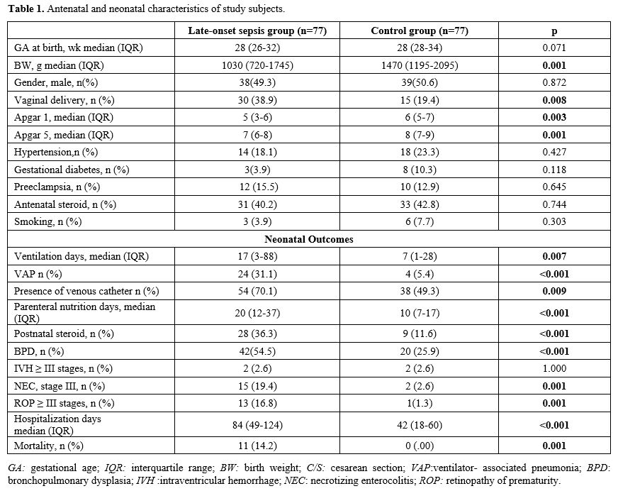
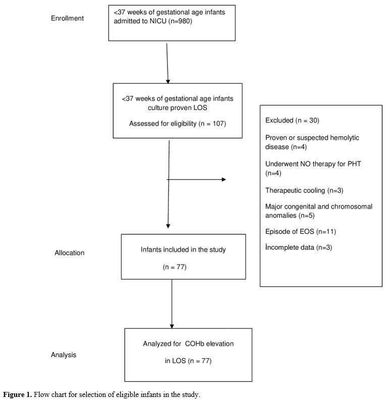
Participants. All infants that were <37 weeks of gestational age born during the study period required NICU admission and were evaluated for inclusion in the study. The LOS group included neonates with culture-proven sepsis after 72h of life. The control group included preterm infants born in our hospital during the same period who met the inclusion criteria and were infection free. In addition, neonates with any of the following were excluded: (1) proven or suspected hemolytic disease, (2) pulmonary hypertension requiring NO therapy, (3) use of therapeutic cooling, (4) presence of major congenital and chromosomal anomalies, (5) incomplete laboratory data or any episode of early-onset sepsis (Figure 1).


Figure 1
Figure1. Flow chart for selection of eligible infants in the study.

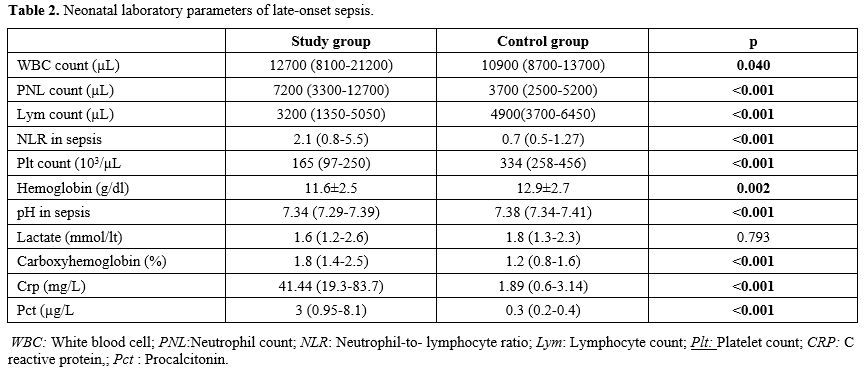
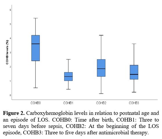
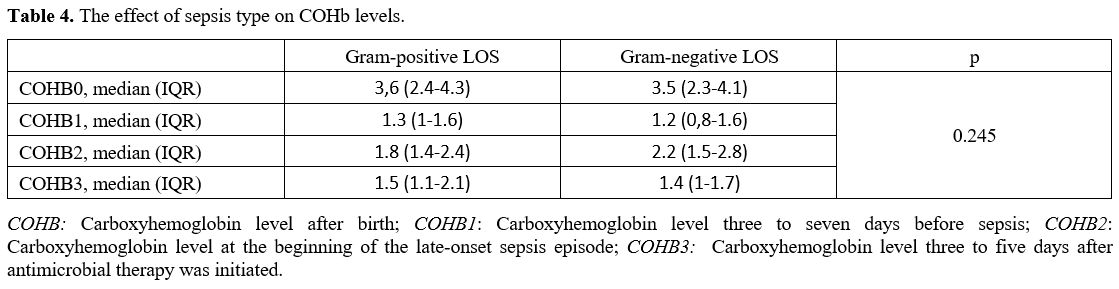
Data collection and definitions. Data were extracted from patients' records. Demographic, perinatal, and neonatal characteristics were recorded and analyzed. Positive blood culture was a prerequisite for LOS. Two positive blood cultures were required for the diagnosis of coagulase-negative staphylococci sepsis. A fully automated BACTEC method (BACT/ALERT 3D system, bioMerieux, SA, France) was used to analyze blood culture isolates. The blood gas analyzer (ABL 800 FLEX, Radiometer, Copenhagen, Denmark) used for the measurement of COHb was calibrated daily. COHb levels were determined from blood sampling that was performed using custom-prepared heparinized containers (Clinitubes, Radiometer, Copenhagen, Denmark), as only a small amount of blood was required (35-55µl). COHb levels are a percentage (%) of total hemoglobin levels. Four-time points were used for assessing COHb levels in the LOS group: one during NICU admission after birth (COHB0); one three to seven days before sepsis (COHB1); one at the beginning of the LOS episode (6h before or 24h after blood culture samples were obtained in the presence of clinical signs) (COHB2); and one three to five days after antimicrobial therapy was initiated (COHB3) (Treatment group). Since the median day for the onset of sepsis was 19 in the LOS group, the COHb level in the control group was assessed to coincide with the first day of the septic attack.
Neonatal morbidities included: intraventricular hemorrhage (IVH), necrotizing enterocolitis (NEC), bronchopulmonary dysplasia (BPD), and retinopathy of prematurity (ROP). Severe IVH included stages ≥ III, classified according to Volpe's cranial ultrasound classification.[8] BPD was defined according to oxygen requirement and ventilator support at 36 weeks of gestation.[9] Severe ROP included stages ≥3 or any stage requiring cryotherapy or laser photocoagulation as classified by standardized international criteria.[10] Bell criteria were used to define severe NEC.[11]
Outcome Measures. The primary outcome measure was the change in COHb levels. Secondary outcome measurements included the presence of a venous catheter, ventilator-associated pneumonia (VAP), postnatal steroids, parenteral nutrition days, duration of ventilation, BPD, NEC, ROP, IVH, hospitalization days, and mortality.
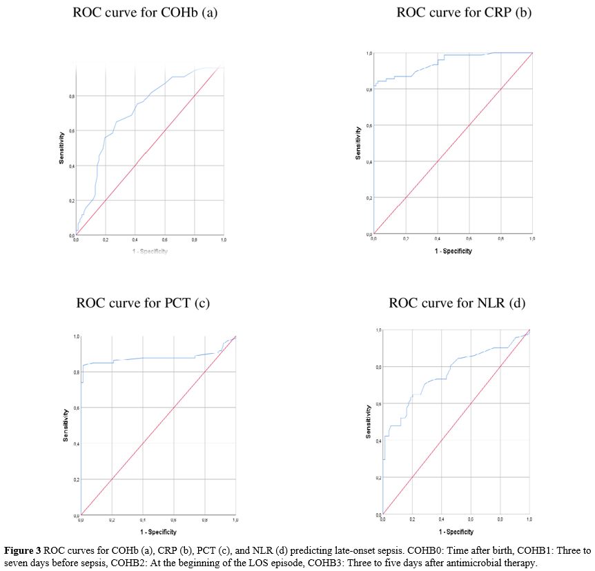
Statistical analysis. "IBM SPSS Statistics for Windows" (IBM Corp. Released 2017, Version 25.0. Armonk, NY, USA) was used for statistical analysis, and "G*Power" version 3.1.9.4 was used to compute the sample size. The sample size was calculated as 142 when an alpha of 0.1, an effect size of 0.432, and a power of 0.90 were used. The Pearson chi-square test was used to compare categorical variables reported as n (%). Continuous variables were expressed using the mean, standard deviation (SD), or medians and ranges (min-max). The normality of data for continuous variables was tested with the Kolmogorov-Smirnov test and compared with either unpaired Student's t-test or the Mann-Whitney U test. The Spearman analysis evaluated the correlation between continuous variables. Repeated Measures Analysis was used to compare the variation in multiple measured values between groups. Receiver operating characteristic (ROC) analysis was used to investigate the COHb values in predicting LOS. The sensitivity, specificity, positive likelihood ratio (+LR), and negative likelihood ratio (-LR) ratio were calculated for the ideal cut-off value. Cut-off values showed the highest sensitivity. Statistical significance was considered when p<0.05 for all tests.

Results

One thousand nine hundred thirty-three infants were admitted to the NICU during the study period; 980 were preterm. Culture-proven LOS has been detected in 11.3% (77/680) of infants. A total of 154 infants met the inclusion criteria of the study. The LOS group consisted of 77 newborns with an average gestation week and birth weight of 28 (26-32) weeks and 1030 (720-1745) gr, respectively. The onset time of LOS, calculated as the median, was 19 (9-42) days in the study group. The control group participants yielded 77 preterm infants with an average gestation week and birth weight of 28 (28-34) weeks 1470 (1195-2095) gr, respectively.
Antenatal and neonatal characteristics of participants in the LOS group are shown in Table 1. When LOS and control groups were compared, spontaneous vaginal delivery (p=0.008), presence of venous catheter (p=0.009), VAP (p<0.001), postnatal steroid use (p<0.001), BPD (p<0.001, mortality (p =0.001), NEC stage III (p=0.001), severe ROP (p=0.001), duration of ventilation (p=0.007), parenteral nutrition (p<0.001) and hospitalization days (p<0.001) were significantly higher in the LOS group.


Table 1
Table 1. Antenatal and neonatal characteristics of study subjects.

Apgar scores were significantly lower in the first (p=0.003) and fifth minute (p=0.001) in the LOS group compared to the control group.
Table 2 demonstrates neonatal laboratory parameters of LOS in all neonates. When the two groups were compared, WBC (p=0.040), PNL (p<0.001), and NLR (p<0.001). COHb (p<0.001), CRP (p<0.001), and PCT (p<0.001) were significantly higher in the LOS group, and the lymphocyte count (p<0.001), platelet count (p<0.001), hemoglobin (Hb) levels (p=0.002), and pH levels (p<0.001) were significantly lower when compared to the control group.


Table 2
Table 2. Neonatal laboratory parameters of late-onset sepsis.

COHb levels were observed to be increased at birth and gradually decreased [3.6 (2.4-4.2) vs.1.5 (1-2)], respectively. However, when an attack of LOS was seen, LOS led to a dramatic rise followed by a decrease after the initiation of the antimicrobial therapy [1.8 (1.4-2.5)] vs. [1.45 (0.2-4)] p<0.001 (Figure 2).

Figure 2
Figure 2. Carboxyhemoglobin levels in relation to postnatal age and an episode of LOS. COHB0: Time after birth, COHB1: Three to seven days before sepsis, COHB2: At the beginning of the LOS episode, COHB3: Three to five days after antimicrobial therapy.

Receiver operating characteristic (ROC) analysis was utilized to establish the best COHb cut-off value (Figure 3). ROC analysis yielded an AUC of 0.714 for COHb (95% CI: 0.631-0.796, p<0.001). At an optimal cut-off of >1.5%, the sensitivity of the test was 64.9%, the specificity was 72.7%, the positive predictive value (PPV) was 70.4%, and the negative predictive value (NPV) was 67.4%. Sensitivity, specificity levels, PPV, and NPV for the other biomarkers are shown in Table 3.

Figure 3 Figure 3. ROC curves for COHb (a), CRP (b), PCT (c), and NLR (d) predicting late-onset sepsis. COHB0: Time after birth, COHB1: Three to seven days before sepsis, COHB2: At the beginning of the LOS episode, COHB3: Three to five days after antimicrobial therapy.

Table 3 Table 3. Comparison of carboxyhemoglobin level with other biomarkers during late-onset sepsis episode.

The ROC analysis found no relation between BPD, mortality, and COHb levels. There was also no correlation between COHb levels and the severity of IVH (r=-0.076; p=0.511), NEC (r=0.003, p=0.980), ROP (r=0.008, p=0.947) and serum lactate (r=-0.057; p=0.485). COHb was discovered to be weakly associated with serum pH (r=0.256, p<0.001).
Gram-positive microorganisms led to more LOS when compared to (53/77, 68.8%) gram-negative microorganisms (20/77, 25.9%). LOS was caused by both gram-positive and gram-negative microorganisms simultaneously in 2 (2/77, 2.60%) and by fungal infections in 2 neonates (2/77, 2.60%). Coagulase-negative staphylococci (CoNS) were the most commonly observed gram-positive organisms that caused LOS (n=37, 67.2%), followed by Staphylococcus aureus (n=11, 20%). Gram-negative microorganism types were as follows: Klebsiella spp. (n=6, 26%), Stenotrophomonas maltophilia (n=4, 17.4%), Serratia spp.(n=3, 13%), Enterobacter spp. (n=3, 13%), Acinetobacter spp. (n=2, 8.7%), Pseudomonas spp. (n=2, 8.7%), Escherichia coli (n=2, 8.7%) and others (n=1, 4.3%). Overall, the positive urine culture rate and cerebrospinal fluid culture rate were 15.7% (n=12) and 8.1% (n=6), respectively. The type of sepsis (gram-positive or gram-negative) did not significantly affect COHb levels [1.8 (1.4-2.4) vs.2.2 (1.5-2.8)] (p=0.245)] (Table 4).

Table 4
Table 4. The effect of sepsis type on COHb levels.


Discussion

Despite the recent significant improvement in neonatal and perinatal care over the last few decades, LOS continues to play an essential role in preterm infant mortality and morbidity. Preterm infants are three to ten times more likely to have an infection when compared to full-term infants due to the immune dysfunction and absence of transplacental immunoglobulin G transmission.[12] According to the literature, prolonged hospitalization period, the need for ventilation devices, central venous access, and the prolonged time spent on total parenteral nutrition before switching to full-enteral feeding were the numerous risk factors for preterm infants to develop LOS in our study.[2] In addition, gram-positive microorganisms are far more prevalent than gram-negative organisms in LOS observed in NICUs.[13] However, we found that the causative microorganism for LOS did not predict COHb elevation.
The challenge in diagnosing LOS persists since no single test with high sensitivity and specificity can quickly and accurately exclude that LOS has been developed. Commonly used biomarkers for evaluating sepsis risk have included CRP, PCT, and complete blood count with increased or decreased leukocyte count, decreased platelet count and increased NLR.[14] Despite its limitations, CRP is commonly used in the diagnosis of sepsis. It may not be elevated if taken early. It is non-specific and may increase in other pro-inflammatory conditions such as NEC. It also may behave differently in preterm and full-term infants.[15] In a meta-analysis of 10 trials, CRP's median sensitivity and specificity were reported to be 70% and 89%.[16] In our study, CRP's median sensitivity and specificity were 84% and 97% higher, respectively.
PCT is another biomarker for diagnosing sepsis. Its secretion starts after two hours of stimulation and peaks at 12-24h with a half-life of about 24h.[17] However, PCT's median sensitivity and specificity were reported to be 85% and 54% in a meta-analysis of 17 trials.[18] Our current work determined PCT's median sensitivity and specificity as 83% and 98%, respectively. Different cut-off points of NLR for LOS in preterm infants have been described, and it is suggested to be used in conjunction with CRP. The sensitivity and specificity of NLR were reported at 83.9% and 79%.[19] However, it was found to be 63% and 79% in our study, respectively. The differences between these markers' sensitivity and specificity rate reported in the literature and our study may be due to differences in populations, including healthy infants, preterm neonates, low birth weight infants, heterogeneities of the sepsis onset time, high-risk factors including intracranial hemorrhage, neonatal hemolysis or perinatal asphyxia. No complete blood count index, such as high or low white blood cell counts, elevated absolute neutrophil counts, or low platelet counts, has demonstrated adequate sensitivity to rule out LOS in the neonatal period.[20] As a result, our study investigated COHb levels as a biomarker in LOS.
There are conflicting and limited results in the literature regarding the use of COHb in LOS in preterm infants. For example, Guney et al. reported that a COHb value of ≥1.35 had a sensitivity of 56.07% and specificity of 90% as a marker of LOS[21] On the contrary, a study on preterm infants did not demonstrate increased COHb levels in sepsis.[22] Our current work shows that a cut-off value of >1.5% for COHb has a sensitivity of 64.94% and a specificity of 72.73%. Although this can be perceived as low when compared to CRP (84.42% and 97.40%) and PCT (83.56% and 98%), it cannot be considered to be too far from NLR (63.38% and 79.73%). Herein, we have shown that COHb levels increase and subsequently decrease with treatment in LOS.
The heme oxygenase (HO) enzyme degrades heme, forming carbon monoxide, iron, and biliverdin. HO has three isoforms: HO-1, HO-2, and HO-3. The gene expression of the first isoform is strongly associated with oxidative stress stimuli such as bacterial lipopolysaccharides, hyperoxia, hypoxia, heat shock, ischemia, UV radiation, H2O2, cytokines, nitric oxide, and heavy metals.[23] Erythrocytes are more vulnerable to oxidative stress in the neonatal period than in adulthood.[24] Fetal hemoglobin has a lower carbon monoxide (CO) binding affinity than hemoglobin A.[25]
Contrary to expectations, the carbon monoxide shift from the placental to maternal circulation leads to lower fetal COHb levels during pregnancy. In addition, the sudden change in oxygenation after birth from low to high oxygen levels promotes HO-1, leading to CO generation.[22] Therefore, COHb levels are high at birth, decreasing gradually and rising again with a LOS attack, as found in our study. Maroti et al. reported that the HO-1 enzyme level increases after birth and falls in term and preterm infants' first week of life.[26] The onset of a LOS attack was a median of 19 (9-42) days in our study. Therefore, the level of COHb can be considered stable at the beginning of the LOS attack in our study. However, considering the variability of COHb level, especially in the postnatal one week, further studies concerning the change over time for the ideal cut-off value for COHb may be needed.
Since hypoxia and low pH have been demonstrated to increase CO dissociation and acidosis to induce HO-1 synthesis, we discovered a negative link between pH and COHb levels [27] Although pH and COHb levels were found to be weakly correlated in our study, the fact that preterm infants with sepsis had lower Apgar scores, weight, longer time spent on total parenteral nutrition, and ventilation may also have contributed to this situation. There are conflicting results in critically ill adult patients regarding a correlation between serum lactate and COHb in the literature.[28,29] However, we found no correlation between serum lactate and COHb in the present study. In an experimental animal model, intestinal HO-1 expression increased in NEC. In this case, increased CO formation has been reported to reduce hypoxia and formula-feeding-induced intestinal inflammation and mortality.[30] However, we found no correlation between COHb levels and the severity of NEC and mortality.
There are several limitations of this study. First, due to the study's retrospective design, we had no control over the timing of samples and ambient oxidative stress variables in the NICU. However, as all infants remained in the same setting, the oxidative stress factors were the same across the board. Another restriction is that more precise technologies exist for measuring COHb levels than co-oximeters. However, none of these methods are practical and require sophisticated equipment. Moreover, we do not have a suspected sepsis group, except for proven sepsis and control groups, which may have caused the biomarkers to show exaggerated performance.
Our study is not the first o to evaluate COHb levels in preterm infants with sepsis. Furthermore, there are conflicting and limited results in the literature regarding the increase of COHb in LOS in preterm infants. However, as far as we know, no previous study has compared COHb levels with other sepsis biomarkers, such as CRP, PCT, and NLR, for diagnosing LOS in preterm infants, more frequently utilized.[15-18,31] We found that the COHb diagnostic value was low compared with CRP and PCT. However, the variations in COHb level in the blood gas analysis, frequently used in preterm infants in the NICU, can be used as a guide when associated with clinical parameters and other biomarkers in diagnosing LOS. Furthermore, new studies with larger sizes can clarify the gaps, such as the validated cut-off for its utility as a bedside test.


Conclusions

In conjunction with other sepsis indicators, the variations of serum COHb levels could be used as a point-of-care test to diagnose LOS in preterm neonates. We have demonstrated that COHb levels may be utilized while diagnosing and treating LOS in NICU. Whether the diagnostic value increases if combined with other biomarkers needs to be studied in prospective trials.


References   

  1. Shane AL, Stoll BJ. Recent developments and current issues in the epidemiology, diagnosis, and management of bacterial and fungal neonatal sepsis. Am J Perinatol. 2013;30(2):131-41.  https://doi.org/10.1055/s-0032-1333413 PMid:23297182
  2. Ran NC, van den Hoogen A, Hemels MAC. Gram-negative Late-onset Sepsis in Extremely Low Birth Weight Infants Is Emerging in The Netherlands Despite Quality Improvement Programs and Antibiotic Stewardship!Pediatr Infect Dis J. 2019;38(9):952-957. https://doi.org/10.1097/INF.0000000000002408 PMid:31274834
  3. McArdle AJ, Webbe J, Sim K, Parrish G, Hoggart C, Wang Y, Kroll JS, Godambe S, Cunnington AJ. Determinants of Carboxyhemoglobin Levels and Relationship with Sepsis in a Retrospective Cohort of Preterm Neonates. PLoS One. 2016;11(8):e0161784. https://doi.org/10.1371/journal.pone.0161784 PMid:27552216 PMCid:PMC4995038
  4. Shi Y, Pan F, Li H, Pan J, Qin S, Jiang D, Shen C. Carbon monoxide concentrations in paediatric sepsis syndrome Arch Dis Child. 2003;88(10):889-90. https://doi.org/10.1136/adc.88.10.889 PMid:14500308 PMCid:PMC1719314
  5. Zegdi R, Perrin D, Burdin M, Boiteau R, Tenaillon A. Increased endogenous carbon monoxide production in severe sepsis. Intensive Care Med. 2002;28(6):793-6. https://doi.org/10.1007/s00134-002-1269-7 PMid:12107688
  6. Turgut K, Yavuz E. Comparison of non-invasive CPAP with mask use in carbon monoxide poisoning Am J Emerg Med. 2020;38(7):1454-1457. https://doi.org/10.1016/j.ajem.2020.04.050 PMid:32331960
  7. Panda SK, Nayak MK, Rath S, Das P. The Utility of the Neutrophil-Lymphocyte Ratio as an Early Diagnostic Marker in Neonatal Sepsis. Cureus. 2021;13(1):e12891. https://doi.org/10.7759/cureus.12891
  8. Inder TE Perlman JM, Volpe JJ. Preterm Intraventricular Hemorrhage/Posthemorrhagic Hydrocephalus. In: Volpe JJ (Ed) Volpe's Neurology of the Newborn 6th Ed. Philadelphia, Elsevier.2018; 637-98. https://doi.org/10.1016/B978-0-323-42876-7.00024-7 
  9. Jobe AH, Bancalari E. Bronchopulmonary dysplasia. Am J Respir Crit Care Med. 2001; 163:1723-9. https://doi.org/10.1164/ajrccm.163.7.2011060 PMid:11401896
  10. International Committee for the Classification of Retinopathy of Prematurity, The international classification of retinopathy of prematurity revisited. Arch Ophthalmol. 2005;123(7):991-9. https://doi.org/10.1001/archopht.123.7.991 PMid:16009843
  11. Pergialiotis V, Konstantopoulos P, Karampetsou N, Koutaki D, Gkioka E, Perrea DN, Papantoniou N. Calprotectin levels in necrotizing enterocolitis: a systematic review of the literature. Inflamm Res. 2016; 65:847-852. https://doi.org/10.1007/s00011-016-0963-9 PMid:27328832
  12. Shane AL, Sánchez PJ, Stoll BJ. Neonatal sepsis. Lancet. 2017;390(10104):1770-1780. https://doi.org/10.1016/S0140-6736(17)31002-4 PMid:28434651
  13. Acheampong EN, Tsiase JA, Afriyie DK, Amponsah SK. Neonatal Sepsis in a Resource-Limited Setting: Causative Microorganisms and Antimicrobial Susceptibility Profile. Interdiscip Perspect Infect Dis. 2022 ;2022:7905727. https://doi.org/10.1155/2022/7905727 PMid:35669534 PMCid:PMC9166966
  14. Goldberg O, Amitai N, Chodick G, Bromiker R, Scheuerman O, Ben-Zvi H, Klinger G. Can we improve early identification of neonatal late-onset sepsis? A validated prediction model. J Perinatol. 2020;40(9):1315-1322. https://doi.org/10.1038/s41372-020-0649-6  PMid:32203177
  15. Eschborn S, Weitkamp JH. Procalcitonin versus C-reactive protein: review of kinetics and performance for diagnosis of neonatal sepsis. J Perinatol. 2019;39(7):893-903. https://doi.org/10.1038/s41372-019-0363-4 PMid:30926891
  16. Liu Y, Zhao L, Wu Z. Accuracy of C-Reactive Protein Test for Neonatal Septicemia: A Diagnostic Meta-Analysis. Med Sci Monit. 2019; 25:4076-4081. https://doi.org/10.12659/MSM.916968 PMid:31152537 PMCid:PMC6559181
  17. Reinhart K, Meisner M, Brunkhorst FM. Markers for sepsis diagnosis: what is useful? Crit Care Clin. 2006;22(3):503-19, ix-x. https://doi.org/10.1016/j.ccc.2006.03.003 PMid:16893736
  18. Pontrelli G, De Crescenzo F, Buzzetti R, Jenkner A, Balduzzi S, Calò Carducci F, Amodio D, De Luca M, Chiurchiù S, Davies EH, Copponi G, Simonetti A, Ferretti E, Di Franco V, Rasi V, Della Corte M, Gramatica L, Ciabattini M, Livadiotti S, Rossi P. Accuracy of serum procalcitonin for the diagnosis of sepsis in neonates and children with systemic inflammatory syndrome: a meta-analysis. BMC Infect Dis. 2017; 17(1):302. https://doi.org/10.1186/s12879-017-2396-7  PMid:28438138 PMCid:PMC5404674
  19. Alkan Ozdemir S, Arun Ozer E, Ilhan O, Sutcuoglu S. Can neutrophil to lymphocyte ratio predict late-onset sepsis in preterm infants? J Clin Lab Anal. 2018;32(4):e22338. https://doi.org/10.1002/jcla.22338 PMid:29055117 PMCid:PMC6817131
  20. Hornik CP, Benjamin DK, Becker KC, Benjamin DK Jr, Li J, Clark RH, Cohen-Wolkowiez M, Smith PB.Use of the complete blood cell count in late-onset neonatal sepsis. Pediatr Infect Dis J. 2012; 31(8):803-7. https://doi.org/10.1097/INF.0b013e31825691e4 PMid:22531232 PMCid:PMC3399981
  21. Guney Varal I, Dogan P. Serial Carboxyhemoglobin Levels and Its Relationship with Late Onset Sepsis in Preterm Infants: An Observational Cohort Study. Fetal Pediatr Pathol. 2020; 39(2):145-155.  https://doi.org/10.1080/15513815.2019.1652377 PMid:31429384
  22. McArdle AJ, Webbe J, Sim K, Parrish G, Hoggart C, Wang Y, et al. Determinants of Carboxyhemoglobin Levels and Relationship with Sepsis in a Retrospective Cohort of Preterm Neonates. PLoS One. 2016;11(8):e0161784. https://doi.org/10.1371/journal.pone.0161784  PMid:27552216 PMCid:PMC4995038
  23. Wagener FA, Volk HD, Willis D, Abraham NG, Soares MP, Adema GJ, Figdor CG. Different faces of the heme-heme oxygenase system in inflammation. Pharmacol Rev. 2003;55(3):551-71. https://doi.org/10.1124/pr.55.3.5  PMid:12869663
  24. Kondo M, Itoh S, Kusaka T, Imai T, Isobe K, Onishi S. The ability of neonatal and maternal erythrocytes to produce reactive oxygen species in response to oxidative stress. Early Hum Dev. 2002;66(2):81-8. https://doi.org/10.1016/S0378-3782(01)00234-1 PMid:11872312
  25. Engel RR, Rodkey FL, O'Neal JD, Collison HA. Relative affinity of human fetal hemoglobin for carbon monoxide and oxygen. Blood 1969;33(1):37-45. https://doi.org/10.1182/blood.V33.1.37.37 PMid:5763632
  26. Maróti Z, Katona M, Orvos H, Németh I, Farkas I, Túri S.Heme oxygenase-1 expression in premature and mature neonates during the first week of life.Eur J Pediatr. 2007;166(10):1033-8. https://doi.org/10.1007/s00431-006-0375-x PMid:17203280
  27. Sharma VS, Schmidt MR, Ranney HM. Dissociation of CO from carboxyhemoglobin. J Biol Chem1976 ;251(14):4267-72. https://doi.org/10.1016/S0021-9258(17)33291-X  PMid:6474
  28. Sedlacek M, Halpern NA, Uribarri J. Carboxyhemoglobin and lactate levels do not correlate in critically ill patients. Am J Ther 1999;6(5):241-4. https://doi.org/10.1097/00045391-199909000-00003  PMid:11329105
  29. Grigorescu BL, Săplăcan I, Bordea IR, Petrisor M, Coman O, Puiac CI, Toncean A, Fodor RS. Endogenous Carboxyhemoglobin Level Variation in COVID-19 and Bacterial Sepsis: A Novel Approach? Microorganisms. 2022;10(2):305. https://doi.org/10.3390/microorganisms10020305 PMid:35208760 PMCid:PMC8878399
  30. Zuckerbraun BS, Otterbein LE, Boyle P, Jaffe R, Upperman J, Zamora R, Ford HR. Carbon monoxide protects against the development of experimental necrotizing enterocolitis. Am J Physiol Gastrointest Liver Physiol. 2005;289(3):G607-13. https://doi.org/10.1152/ajpgi.00055.2005 PMid:15890710
  31. James J., Tewari V.V., Jain N. Diagnostic accuracy of clinical tool 'STOPS' and serum procalcitonin for optimizing antibiotic therapy in neonates born at ≥28 weeks of gestation with neonatal sepsis. Mediterr J Hematol Infect Dis 2021, 13(1): e2021019 https://doi.org/10.4084/mjhid.2021.019 PMid:33747400 PMCid:PMC7938925

[TOP]