Liver Disease and Sickle Cell Disease: Auto-Immune Hepatitis more than a Coincidence; A Systematic Review of The Literature

Kelvin Lynch1*, Andrea Mega2*, Massimo Daves3, Asma Sadiq4, Helen Fogarty5,6 and Andrea Piccin4,7,8.

1 Dept of Gastroenterology, Cork University Hospital, Cork, Ireland.
2 Dept of Gastroenterology, Provincial Hospital of Bolzano (SABES-ASDAA), Bolzano, Italy.
3 Dept of Laboratory Medicine, Provincial Hospital of Bolzano (SABES-ASDAA), Bolzano, Italy.
4 Northern Ireland Blood Transfusion Service, Belfast, UK.
5 Department of Haematology, Children’s Health Ireland at Crumlin, Dublin.
6 Irish Centre for Vascular Biology, Royal College of Surgeons in Ireland, Dublin.
7 Dept of Internal Medicine V, Medical University of Innsbruck, Innsbruck, Austria.
8 Dept of Industrial Engineering, University of Trento, Trento, Italy.

Correspondence to: Prof. Andrea Piccin,  Consultant Haematologist, FRCPath, PhD, MD.  Northern Ireland Blood Transfusion Service, Belfast, UK. E-mail: andrea.piccin@nibts.nhsni.net

Published: November 1, 2023
Received: June 27, 2023
Accepted: October 14, 2023
Mediterr J Hematol Infect Dis 2023, 15(1): e2023060 DOI 10.4084/MJHID.2023.060

This is an Open Access article distributed under the terms of the Creative Commons Attribution License
(
https://creativecommons.org/licenses/by-nc/4.0), which permits unrestricted use, distribution, and reproduction in any medium, provided the original work is properly cited.

Abstract

In patients with SCD, chronic liver damage is a common manifestation. More than 50% of SCD patients have elevated liver enzymes. Common underlying aetiologies include sickle cell hepatic crisis, viral hepatitis, sickle cell intrahepatic cholestasis and hepatic sequestration in the acute setting, and cholelithiasis and iron overload in the chronic setting. Autoimmune hepatitis (AIH) is a rare disease that appears to occur more commonly in the sickle cell disease (SCD) population than in the general population. There are many schools of thought as to why this is the case, including the phosphatidylserine hypothesis, the heme inflammatory hypothesis, the complement generation hypothesis, and the transfusion alloimmunization hypothesis.
Due to the natural history of the two illnesses, SCD is almost always diagnosed first in cases of dual pathology. Symptoms such as jaundice, fatigue, and abdominal pain are common in SCD, as are abnormal liver function tests (LFTs). These abnormalities, attributed to the other more frequent liver involvements in SCD, can lead to delays in AIH diagnosis in this population.
Corticosteroids, sometimes with other immunosuppressive agents, such as azathioprine, are the cornerstone of acute AIH treatment (). However, corticosteroid use in the SCD population has been shown to carry an increased risk of vaso-occlusive crises, providing a treatment dilemma.
The following is a review of AIH in the SCD population, where we explore the pathophysiology behind the association between the two disorders, discuss an approach to investigating abnormal LFTs in SCD, and examine treatment options in this population with co-existing diseases.


Introduction

Sickle cell disease (SCD) is a genetic disorder which occurs because of polymerisation of the haemoglobin molecule resulting in increased erythrocyte adhesion with subsequent vaso-occlusion as well as haemolysis.[1] Its prevalence ranges among different ethnic groups with approximately 300,000 children being born each year worldwide with sickle cell anaemia (SCA) (HbSS).[2]
In patients with SCD, chronic liver damage is a common manifestation. One study prospectively reviewed 170 SCD patients to find 69% had elevated liver enzymes.[3] Common underlying aetiologies include sickle cell hepatic crisis, viral hepatitis, sickle cell intrahepatic cholestasis and hepatic sequestration in the acute setting, and cholelithiasis and iron overload in the chronic setting.[4,5] These can lead to clinical and biochemical abnormalities in patients, including jaundice, hepatomegaly and deranged liver function tests.
The sickling process favours hyperbiluribinaemia with gallstone formation, hypoxic liver injury and hepatic sequestration.[6] The pathological mechanism of liver damage during SCD has been studied only in post-mortem studies, since liver biopsy is not advised due to the high risk of bleeding and death (up to 28%).[7] The liver damage classically involves the sinusoidal spaces which are obstructed or dilated. The presence of free hemoglobin and free iron will damage the endothelial cells favouring a vicious inflammatory circle.[8] The obstruction of the sinusoid could lead to infarcts and even necrosis of the centrilobular area, followed by fibrosis. The Kupfer cell are usually expanded and erthrophagocytosis is present. Over time, all these processes may favour the formation of peculiar nodular inclusions (Gamna Gandy Bodies) and liver-iron overload.[9] Hepatic iron overload is also a consequence of transfusion therapy in SCD, especially if iron chelation is not performed or not well monitored.[10] Non-transferrin bound iron (NTBI) will deposit in the liver parenchima and facilitate the generation of reactive oxygen species (ROS). ROS will further sustain hepatic cellular damage.
Viral Hepatitis mostly derived from blood transfusions is an important cause to consider when SCD patients have deranged liver disfunction, particularly in countries where the risk of Transfusion Transmitted infection (TTI) is still very high.[11] In Oman, amongst a total of 1000 SCD patients (491 males and 509 females), twenty-three (2.3%) patients showed positive serology for the hepatitis B virus (HBV) surface antigen (HbsAg), of whom sixteen (1.6%) were HBV DNA positive. 126 (12.6%) had anti-Hepatitis C virus (HCV) antibodies (anti-HCV), of whom fifty-two (5.2%) were HCV RNA positive.[12] In Senegal, the prevalence of HCV in SCD-transfused was 1.33% and HBV prevalence was 2%.[13] Viral hepatitis A (HAV) can also lead to fulminant hepatitis in the SCD population.[14] Prophylactic vaccinations against HAV and HBV should always be encouraged. Tuberculosis could also affect the liver of patients with SCD.[15]
There are many established common complications of SCD, one of which is vaso-occlusive crises. This is characterised by extreme pain and often requires hospital admission with intravenous opiate analgesia.[16] In more severe cases exchange transfusion may be needed.[17] Low molecular weight heparin has been shown to reduce the duration of these crises.[18] Many precipitating factors for vaso-occlusion exist, including the use of corticosteroids.[19]
An increased risk of developing auto-immune disease in SCD has been reported.[20] Recently there has been a dramatic increase in the number of SCD patients living in Western countries.[21] Hence the prevalence of co-existing conditions among this population, including liver conditions is also likely to increase year-on year and an awareness of how best to manage these co-morbidities is essential.
Many aetiologies of auto-immune liver disease (AILD) exist with autoimmune hepatitis (AIH), primary sclerosing cholangitis (PSC) and primary biliary cholangitis (PBC) being common subtypes. AIH is a subtype with good evidence for the use of corticosteroid when treating.[22] It is characterized by the presence of circulating non-organ and liver-specific autoantibodies and is usually responsive to immunosuppression.[23] It is thought to occur due to a combination of genetic predisposition, environmental triggers and a failure of the immune response.[24] AIH has a reported prevalence of approximately 31/100,000[25] and is usually seen in adults and only very rarely in children.
The increased risk of auto-immunity in SCD, the potential for clinical and biochemical abnormalities of undiagnosed AILD being attributed to existing SCD, and the possible side effect of vaso-occlusive crises when AIH is treated with corticosteroid in a SCD patient, has prompted us to conduct a systematic review of the relevant literature to assess the prevalence of AIH and other AILDs in the SCD population. We also looked at the diagnostic challenge of AILD in SCD and the therapeutic options for the patients in whom AIH and SCD co-exist.
A systematic review was performed on PubMed using the words “sickle cell anemia” and ”hepatitis”, this gave an original list of 294 publications (updated to the 31.08.2023). Out of these 21 were selected that also matched the word “autoimmune”.

Prevalence of AILD in SCD

AILD is one of the various liver diseases present in drepanocytosis and is certainly not the most frequent cause. The most frequent causes in the countries where this disease is most widespread and are linked to transfusion treatment and are viral hepatitis and martial overload. However, it was noted by Jitraruch et al that 13 of the 77 (17%) patients with SCD who presented to their centre with hepatobiliary complications, had an autoimmune cause as the underlying aetiology.[19] Elsewhere, Li-Thiao-Te et al reported 2 cases of AIH in their population of 603 patients.[26] Waisbord-Zinman et al reported a further seven patients, ages 8 to 23 years with AIH, PSC, and AIH/PSC overlap.[27] Often the involvement of the liver in SCD patients is misdiagnosed because jaundice is ascribed to hemolysis, rather than being considered of primary hepatic origin. The exact mechanism for the immune tolerance breakdown in AIH has not been unveiled. However, genetic predisposition, molecular mimicry, and impaired T cell number and function are considered key actors.[28]
Although no strong conclusion can be taken from the limited data available on the prevalence of AILD in the SCD population, these numbers are suggestive that it may be greater than would be expected if they were independent events.

The Role of the Spleen in AILD with SCD

When discussing AILD in SCD, a particular consideration to the role of spleen should be given. Patients with SCD develop over time functional hyposplenism.[29] This is the consequence of repeated vaso-occlusive events within the spleen parenchima. These will destroy the white pulp leading to several degenerative processes including Gamna Gandi body formation.[9] Patients with SCD require therefore lifelong prophylaxis against capsulated bacteria and vaccinations (Meningococcus, Haemophilus influenzae, Streptococcus pneumoniae etc). Small children are prone to develop splenic sequestration which are lifethreatening and in these cases splenectomy is recommended.[30] However, splenectomy has always been a procedure recommended when autoimmune conditions are resistant to treatment, for example in idiopathic thrombocytopenia. For these reasons, we believe that clinicians and researchers should be aware of the possibility that hypofunctional spleen may indirectly prevent AILD manifestation, indirectly protecting patients.

AILD Diagnosis

AILD often manifests as a chronic disease with an insidious onset of chronic liver disease symptoms. The diagnosis is often made after incidental discovery of abnormal liver function tests but sometimes AILD can present as acute liver failure. With regard to AIH specifically, up to 20% of patients present with acute hepatitis.[31]
There are no pathognomonic diagnostic tests for AILDs and diagnosis is usually based on a combination of clinical, serological, biochemical, and histological findings.
Investigation of AILD usually involves a combination of auto-antibody measurement and histology evaluation. Other hepatitis causes including viral hepatitis should also be screened for using serological investigations as summarized below (Table 1). With regard to AIH, there are common antibodies which should be checked (Table 2) and The AIH Group’s criteria should be applied to calculate a score (Table 3).[32] A simplified score of >7 is 95% specific for AIH.[33]


Table 1 
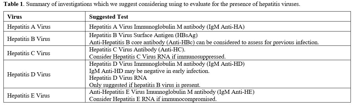
Table 1. Summary of investigations which we suggest considering using to evaluate for the presence of hepatitis viruses.

Table 2 
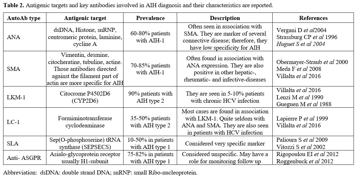
Table 2. Antigenic targets and key antibodies involved in AIH diagnosis and their characteristics are reported.

Table 3 
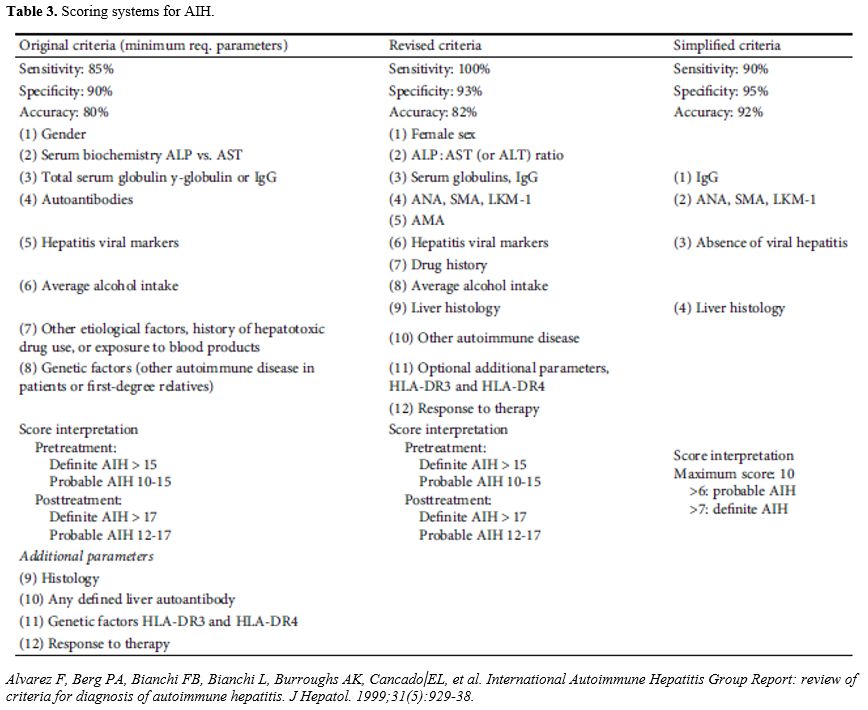
Table 3. Scoring systems for AIH.

PBC and PSC similarly have a multifactorial approach to diagnosis. Radiological evaluation is often useful in addition to autoantibodies and biopsy.[34,35] Overlap syndromes of both AIH and either PBC or PSC (AIH/PBC or AIH/PSC) are more common than expected, if they were independent events. Overlap syndromes tend to be a challenging diagnosis.[36,37] There are special Paris criteria with 97% specificity for AIH/PBC.[38] Unfortunately, AIH/PSC doesn’t have a diagnostic system which is as established. Accurate diagnosis with overlap is important as without an AIH component corticosteroid is not likely to be as therapeutically beneficial and may precipitate vaso-occlusion in patients with co-existing SCD.[26,34,35]


The Diagnostic Challenge

When AILD and SCD co-exist, the diagnosis of SCD almost always pre-dates that of AILD due to the natural history of the diseases.[39-41] For the aforementioned reasons evidence of liver dysfunction is common in the SCD population, however they may also be a sign of new AILD and these should be screened for appropriately. We take AIH as a subsection of AILD to focus on, as accurate diagnosis is vital as steroid therapy is very effective in this disease but can precipitate vaso-occlusion in SCD.[26,34,35]
Before any speculation on co-existing SCD with AIH, we first looked at AIH in the general population. AIH is approximately seven times more common in females than in males and most commonly presents as acute hepatitis. For this diagnosis, serological investigations and the support of a hepatologist for the correct interpretation are important (Table 2). However, it may also present as fulminant hepatitis. Often non-specific symptoms such as arthralgia and fatigue are the first symptoms noted by an otherwise healthy patient.[42] Other common presenting features are jaundice and raised liver function tests (LFTs). A previous Japanese study reported a mean alanine transaminase (ALT) of 776 U/L early during AIH presentations.[43] Many of these presenting features could be attributed to a previously known diagnosis of SCD if one were present.
Raised IgG levels and circulating auto-antibodies are specific indicators of hepatitis of autoimmune aetiology. These autoantibodies consist of Anti-nuclear antibody (ANA), Smooth muscle antibody (SMA), Anti-liver Kidney Microsomal Type 1 (anti-LKM1), Anti-Liver Cytosolic Protein Type 1 (anti-LC1) and more rarely anti-mitochondrial antibody (AMA).[32] Furthermore, auto-Ab testing against soluble liver antigen/liver-pancreas (SLA/LP) have been suggested as useful AIH markers. This is particularly useful for these patients with severe disease.[44]
On the basis of auto-Ab profile, AIH can be categorised in two distinct subtypes:
•    AIH type-1 that is characterized by the presence of ANA and/or SMA.
•    AIH type-2 that is characterized by the presence of LKM-1 and/or LC-1.
The prevalence and the antigenic targets of the principal autoAb seen in AIH are reported in Table 2. Since pathognomic criteria are lacking, AIH is a diagnosis of exclusion that is formulated after out-ruling other causes of chronic hepatitis and according to the criteria from the International Autoimmune Hepatitis Group (Table 3).[32] Furthermore, in 2008 a simplified diagnostic algorithm has been suggested. This tool considers 4 parameters only: i) auto-Ab; ii) serum immunoglobulin; iii) classical histological finding; iv) absence of viral markers.[45]
In SCD, AIH presentation includes: fatigue, arthralgia, and jaundice independent of hepatitis and deranged liver function tests (LFTs). Given that these features are not uncommon in SCD,[46] this can lead to AIH being masked, particularly in the absence of an auto-antibody screen. Positivity of any of the aforementioned auto-antibodies would then be more specific for AIH. Another useful clue in AIH diagnosis within the SCD population is a family history of AIH.
The large crossover of the presenting factors in AILD and chronic SCD symptoms and signs presents a diagnostic challenge. We suggest looking for the specific markers of AIH and other AILDs if there is any new indicators which could be suggestive of a new primary hepatic issue (eg new or worsening deranged liver function tests). It should also be noted that multiple cases of fulminant hepatitis secondary to AIH in the paediatric SCD patients have been reported,[47] suggesting that clinicians should be vigilant for AIH in the SCD population at a young age.


What Triggers Autoimmunity In SCD?

The phosphatidylserine hypothesis. The polymerization process typical of SCA is responsible for damage to the erythrocyte cell membrane leading to hemolysis. Therefore, in SCA the lifespan of red cells is reduced from 120 days to only 10-12 days. These abnormal processes involving the cell membrane favor the generation of microparticles (MP).[48] The exposure of phosphatidylserine on MP and on the external cell membrane of erythrocytes is a strong chemo-active signal enhancing and favoring the recruitment of leukocytes (Figures 1a,1b,1c). This possibly explains why a raised white cell count is documented during sickle cell crises and/or vaso-occlusive events in SCD. Interestingly, this is in accordance with the well-known immunological hypothesis of the “danger model”.[49] Obviously, the degree of sickling is variable over time and can fluctuate. Therefore, patients move from steady state to crises. The sickling mechanism undergoes some thermodynamic rules and can be enhanced or diminished by changes in hydration status, pH and temperature. Nevertheless, a certain degree of sickling with consequent MP generation is always ongoing in SCD. This translates into a constant stimulus for leukocyte recruitment and activation, even during steady state. It is therefore very likely that this continuous activation can make SCD patients more prone to the development of autoimmune disease.[50]

Figure 1a Figure 1A. Normal Erythrocyte. Note the close-up view of the external cell membrane of the erythrocyte and of the haemoglobin molecule of which
the erythrocyte contains multiple.

Figure 1b Figure 1B. Sickle Cell. Note the polymerization of haemoglobin (S) molecules.

Figure 1c Figure 1C. Sickling. Note that microparticles are external to the sickle due to disruption of the cell membrane and are recruiting a leukocyte.

Heme inflammatory hypothesis. The hemolytic process key to the pathogenesis of SCA results in the release of large quantities of free heme into the circulation. This will cause endothelial damage, scavenging of nitric oxide (NO) and also increase the presence of reactive oxygen species (ROS). Free heme also affects leukocyte activation.[51-53] Furthermore, hemolysis induces heme oxygenase 1 (HO-1) which impairs the capacity of leukocytes to destroy pathogenic agents.[54] Therefore, during sickle crises a “storm” occurs within the vessels with swirling of broken red cells, cellular “dust” and MP generation.[50] This “storm” will sustain the exposure of new antigens, and these may trigger inflammatory response, immune activation, and favor autoimmunity.
Complement generation hypothesis. AIlD being more common in the SCD population is further suggested by a theory of increased auto-immunity in SCD. It has been suggested that this is the case due to complement-factor-B consumption in SCD leading to impaired ability to mount a response against infection. This then causes prolonged exposure to antigens which can then trigger an auto-immune response.[20,55] Patients with SCD are polytransfused. The repeated exposure to foreign red cells antigens trigger alloimmunization. Moreover, the presence of a constant inflammatory status, such as in SCD, further potentiates alloimmunization.[56,57] The consequence of alloimmunization is haemolysis mediated by complement. In summary, macrophages bind to erythrocytes coated with IgG, facilitated by macrophage expression of surface Fc receptors specific for the Fc portion of IgG.[57] Any erythrocytes covered by C3b will be removed in the liver by the Kupfer cells.
Interestingly, the liver itself contributes in part to the synthesis of complement, which occurs within the Kupffer cells. Moreover, the red cell membrane alterations typical of SCD will sustain MP production and complement activation.[48,50] Lombardi and colleagues recently reported that the alternative complement pathway is, to a certain degree, always activated in SCA.[58]
Over time, patients with SCA could theoretically develop a consumption of complement (C3b) in a similar fashion to what is reported of the natural occurring anticoagulant molecules: Protein C and Protein S in SCA.[59] A recent study by Gerogianni A and colleagues support this hypothesis. These authors showed that free heme, which is classically increased in SCA, will bind to complement inhibitor factor I and therefore allow C3b to circulate and to cause hemolysis.[60] In addition, the complement components which are not directly involved in the opsonization process, (e.g., C3a, C2a, C5a etc.) can act as anaphylatoxins, enhancing the inflammation and endothelial damage, typical of SCA. Interestingly, the loss of complement regulation as above described, contributes to the development of other autoimmune diseases (e.g., inflammatory arthritis and lupus nephritis).[61]
For all these reasons, cases with SCA and co-existing AIH or other autoimmune disease affecting the liver, present both a clinical challenge and a complex pathogenesis, which merit further scientific investigation.
Transfusion alloimmunization and liver antigens. Kazuaki Tokodai, et al found that ABO antigens are expressed on endothelial cells within the hepatic triad.[62] Earlier studies showed that expression of blood group antigens is common in a normal biliary tree. Moreover, periportal hepatocytes showed neo-expression of blood group antigens in pathologic conditions, (e.g., H substance and even Lea and Leb red blood cells antigens).[63] The possibility that chronic blood transfusion may trigger alloantibodies directed against those antigens cannot be excluded. However, this still needs to be proven.

Management.

The treatment of SCD patients with AILD should be individualized and adapted according to clinical response, with attention to potential side effects. The underlying aetiology of AILD should be treated in accordance with the best evidence. The evidence for using steroids in many AILD including PBC and PSC is limited with ursodeoxycholic acid being the mainstay of treatment in these diseases.[34,35] However, with AIH steroids are the mainstay of treatment which prompts specific considerations in the SCD population, given the concern regarding vaso-occlusion.[64]

AIH Treatment

The treatment of AIH is largely based on the use of corticosteroids and azathioprine (AZA). The goal of AIH treatment is to induce and maintain complete suppression of the inflammatory activity and to prevent disease progression to cirrhosis and liver failure. Remission is achieved when clinical symptoms are absent, and transaminases and immunoglobulins have returned to normal levels. The induction therapy in AIH usually includes a combination of high-dose prednisolone with or without AZA. EASL guidelines furthermore advise that, in patients in whom steroid-specific side effects are expected, remission can also be induced by replacing prednisolone with budesonide at a starting dose of 9 mg/day, but budesonide is ineffective in the presence of cirrhosis.[65]
After a successful 4-week induction therapy in which tapering of steroids has already started, a maintenance phase is usually initiated with continuous fixed doses of 10mg of prednisolone and 50mg of AZA daily, until normalization of serum transaminases, bilirubin, and IgG levels is achieved, and resolution of histological abnormalities becomes evident.[64] AZA treatment is usually continued for at least two years,[66] and subsequent decision to discontinue therapy generally balances the pros of long-term drug-free remission and cons of relapse-risk need for retreatment. Many patients and physicians may consider the risk of relapse with further steroid requirement, unreasonable in the SCD population.
Alternative options to steroid and AZA are also present. Mycofenolate mofetil (MMF) in combination with prednisolone has been shown to be effective and safe in inducing disease, with a recent meta-analysis demonstrating that this combination yielded higher remission rates and lower non-response rates than standard treatment.[67] In a retrospective study on 22 AIH patients with AZA intolerance, 6-MP seemed to be beneficial and tolerated as second-line treatment to AZA.[68] Cyclosporine A has been successfully used as an alternative therapy of AIH patients not responding to AZA and steroids, although the number of patients treated with this strategy is small to date.[69] Early data on mTOR inhibitors indicate successful treatment of refractory AIH and recurrent or de novo post-transplant autoimmune hepatitis in a small number of patients.[70] Biologic options including rituximab and infliximab have also been used in patients with difficult to treat AIH to good effect but again in a small number of patients.[71,72] Tacrolimus, use as frontline treatment in AIH is currently not supported.[73] These are all options which can be considered in sickle cell disease but there is limited data to suggest that there is a non-steroid regimen which is as efficacious as steroids AIH.

AIH Treatment in SCD

Any of the above AIH treatment options can be considered effective when they are able to bring about disease remission. This is considered achieved when: clinical symptoms are absent, and transaminases and immunoglobulins have come to normal levels. The combination of steroids and AZA is as expected the most commonly reported regimen to treat AIH in the SCD population and has been successfully in inducing and maintaining remission in the SCD population.[27,40,41,47]
Unfortunately, steroid therapy can precipitate vaso-occlusive crises in SCD[19,26] and this has been specifically reported in the context of high dose steroids (1.3 mg/kg body weight per day) used for treating AIH in a child with known sickle cell disease.[39] Lower doses of steroid were successful in inducing remission after the vaso-occlusion in this case. This leads to a difficult decision process for a treating physician to balance inducing AIH remission of AIH while avoiding triggering SCD exacerbation. Steroids increase the risk of painful crises as well as stroke.[74]
For these reasons, steroid sparing agents are of utmost importance. AZA is the most commonly reported non-steroid drug used for the treatment of AIH when SCD co-exists. It appears to be successful in maintaining remission in some patients with no major side effects reported.[19,26,41] However, relapse despite AZA was a concern.[39] Suboptimal compliance with AZA was thought to be a factor in these cases. As more data becomes available monoclonal antibodies (eg rituximab) should also be considered.
Outside of the word of liver disease, Valerie Li-Thiao-Te and colleagues successfully used exchange transfusion and hydroxyurea before steroid treatment to reduce the risk of vaso-occlusion in patients with SCD.[26] While exchange transfusion may certainly help reducing the inflammatory status, we believe that the administration of steroids should be carefully evaluated and used when necessary and exchange transfusion should administered only in high dependency units. In many AIH cases, steroids will be necessary and therefore exchange transfusion may be a sensible consideration.
In patients with end stage cirrhotic liver disease secondary to AIH (or other AILDs), liver transplant may be the only valid treatment option. Liver transplantation has been successfully performed in people with co-existing AIH and SCD.[19,26,75] Overall, liver transplant in SCD has been reported to have a 10 year overall survival (OS) of 44.4%.[76] However, this figure includes patients with HCV whom had a high risk of disease reactivation and subsequent death. Careful selection of liver transplant candidates is without doubt imperative but we have evidence to show that orthotopic liver transplant is a good option in patients with SCD and end-stage liver disease.
Calore E and her Italian colleagues from Padova University, as first reported a patient with co-existing AIH and SCD, who underwent a haploidentical stem cell transplant (HSCT).[77] This patient had complete resolution of both their AIH and SCD following a severe initial SCD phenotype. This pioneer case clearly suggest that stem cell transplant could be considered and encouraged in similar cases. Furthermore, the recent availability of monoclonal drugs targeting key players of complement cascade such as anti-C5 (eculizumab®) or the most recent anti Factor D (ALXN2040) may offer new hope for the treatment of AIH and SCD.[61]
When AILD and SCD co-exist, it is important to optimise non-autoimmune contributors of the patient’s liver injury. This includes monitoring iron levels and ensuring adequate iron chelation. However, it is important to note that that hepatic toxicity caused by deferasirox has also been reported.[4,78]

Final Remarks

Early detection of AILD in patients with SCD is important. The most useful factor for diagnosis of ALD in this population is the presence of an autoantibody. We believe that it would be reasonable to perform an auto-antibody screen in any patient with SCD who has a shown unexplained (i.e. not during a crisis episode) elevated liver enzymes. If an AILD is diagnosed, liver imaging and/or transient elastography and/or liver biopsy are recommended.
With regard to AIH specifically, the treatment should include low dose prednisolone +/- AZA if not contraindicated (eg: thiopurine methyl transferase variant). Despite an increased side-effect profile with corticosteroids in SCD, there is no evidence to suggest that an alternative regimen is as effective in inducing remission and Lykavieris and colleagues reported consistent clinical, and biochemical improvement with corticosteroid therapy in patients with histologically confirmed autoimmune hepatitis.[39] For this reason, we would suggest using corticosteroids in this patient cohort. We suggest commencing prednisolone at a low dose of 30mg for an adult patient with AZA being commenced 2 weeks later, usually at a starting dose of 0.5mg/kg but with the potential to titrate upwards. Steroid should be gradually tapered once LFT and IgG levels have normalised. It is recommended that AZA should be continued for at least two years after remission of AIH but in SCD a more prolonged course should be considered.
Patients with SCD are often on treatment with hydroxyurea, for example due to previous severe crises (chest crises, recurrent painful crises etc). This may also have a role in controlling a possible autoimmune driver. Therefore, discontinuing this drug should be carefully evaluated. If a SCD patient is commenced on corticosteroids patients should be strictly monitored, since an increased risk of vaso-occlusion has been reported. Alternative immunosuppressive therapies can be considered, although there is little evidence for these in the SCD population. Liver transplant may ultimately be required and there is evidence to suggest, that a good outcome is possible following transplantation in this cohort, with appropriate patient selection.
In conclusion, although still rare, AILD appears more common in patients with SCD than the general population. It is a challenging diagnosis, as symptoms suggestive of AILD are often already present this population due to their underlying SCD. A positive autoantibody is a key component in obtaining a diagnosis. The treatment of AIH in SCD is complicated further by steroids being shown to precipitate vaso-occlusive crises in some SCD patients. However, no treatment to date has been shown to be as effective in inducing remission of AIH.

References   

  1. Sundd P, Gladwin MT, Novelli EM. Pathophysiology of Sickle Cell Disease. Annu Rev Pathol. 2019;14:263-92. https://doi.org/10.1146/annurev-pathmechdis-012418-012838 PMid:30332562 PMCid:PMC7053558  
  2. Piel FB, Steinberg MH, Rees DC. Sickle Cell Disease. N Engl J Med. 2017;376(16):1561-73. https://doi.org/10.1056/NEJMra1510865 PMid:28423290  
  3. Maher MM, Mansour AH. Study of Chronic Hepatopathy in Patients With Sickle Cell Disease. Gastroenterology Res. 2009;2(6):338-43. https://doi.org/10.4021/gr2009.12.1327 PMid:27990203 PMCid:PMC5139694  
  4. Allali S, Taylor M, Brice J, de Montalembert M. Chronic organ injuries in children with sickle cell disease. Haematologica. 2021;106(6):1535-44. https://doi.org/10.3324/haematol.2020.271353 PMid:33626864 PMCid:PMC8168494
  5. Theocharidou E, Suddle AR. The Liver in Sickle Cell Disease. Clin Liver Dis. 2019;23(2):177-89. https://doi.org/10.1016/j.cld.2018.12.002 PMid:30947870  
  6. Praharaj DL, Anand AC. Sickle Hepatopathy. J Clin Exp Hepatol. 2021;11(1):82-96. https://doi.org/10.1016/j.jceh.2020.08.003 PMid:33679049 PMCid:PMC7897874  
  7. Zakaria N, Knisely A, Portmann B, Mieli-Vergani G, Wendon J, Arya R, et al. Acute sickle cell hepatopathy represents a potential contraindication for percutaneous liver biopsy. Blood. 2003;101(1):101-3. https://doi.org/10.1182/blood-2002-06-1823 PMid:12393551
  8. Piccin A, Murphy C, Eakins E, Rondinelli MB, Daves M, Vecchiato C, et al. Insight into the complex pathophysiology of sickle cell anaemia and possible treatment. Eur J Haematol. 2019;102(4):319-30. https://doi.org/10.1111/ejh.13212 PMid:30664257
  9. Piccin A, Rizkalla H, Smith O, McMahon C, Furlan C, Murphy C, et al. Composition and significance of splenic Gamna-Gandy bodies in sickle cell anemia. Hum Pathol. 2012;43(7):1028-36. https://doi.org/10.1016/j.humpath.2011.08.011 PMid:22079355
  10. Delicou S, Maragkos K, Tambaki M, Kountouras D, Koskinas J. Transient Elastography (TE) is a Useful Tool for Assessing the Response of Liver Iron Chelation in Sickle Cell Disease Patients. Mediterr J Hematol Infect Dis. 2018;10(1):e2018049. https://doi.org/10.4084/mjhid.2018.049 PMid:30210742 PMCid:PMC6131104  
  11. Rezvan H, Abolghassemi H, Kafiabad SA. Transfusion-transmitted infections among multitransfused patients in Iran: a review. Transfus Med. 2007;17(6):425-33. https://doi.org/10.1111/j.1365-3148.2007.00794.x PMid:18067646  
  12. Alkindi S, Al-Umairi N, Jaju S, Pathare A. Prevalence of Hepatitis B, Hepatitis C, and HIV in Multiply Transfused Sickle Cell Disease Patients from Oman. Mediterr J Hematol Infect Dis. 2019;11(1):e2019058. https://doi.org/10.4084/mjhid.2019.058 PMid:31700583 PMCid:PMC6827606  
  13. Seck M, Senghor AB, Loum M, Touré SA, Faye BF, Diallo AB, et al. Transfusion Practice, Post-Transfusion Complications and Risk Factors in Sickle Cell Disease in Senegal, West Africa. Mediterr J Hematol Infect Dis. 2022;14(1):e2022004. https://doi.org/10.4084/MJHID.2022.004 PMid:35070211 PMCid:PMC8746941  
  14. Renge RL, Dani VS, Chitambar SD, Arankalle VA. Fulminant hepatitis A in children with sickle cell disease. Indian Pediatr. 2002;39(2):186-9.  
  15. Droz N, De Lauzanne A, Holvoet L, Missud F, Benkerrou M, Brousse V, et al. Tuberculosis in children with sickle cell anaemia: a retrospective study in French tertiary care centres. Eur J Pediatr. 2017;176(6):723-9. https://doi.org/10.1007/s00431-017-2905-0 PMid:28401342  
  16. Quinn CT, Lee NJ, Shull EP, Ahmad N, Rogers ZR, Buchanan GR. Prediction of adverse outcomes in children with sickle cell anemia: a study of the Dallas Newborn Cohort. Blood. 2008;111(2):544-8. https://doi.org/10.1182/blood-2007-07-100719 PMid:17909076 PMCid:PMC2200853  
  17. Chou ST, Fasano RM. Management of Patients with Sickle Cell Disease Using Transfusion Therapy: Guidelines and Complications. Hematol Oncol Clin North Am. 2016;30(3):591-608. https://doi.org/10.1016/j.hoc.2016.01.011 PMid:27112998  
  18. Robieux IC, Kellner JD, Coppes MJ, Shaw D, Brown E, Good C, et al. Analgesia in children with sickle cell crisis: comparison of intermittent opioids vs. continuous intravenous infusion of morphine and placebo-controlled study of oxygen inhalation. Pediatr Hematol Oncol. 1992;9(4):317-26. https://doi.org/10.3109/08880019209016603 PMid:1467165  
  19. Jitraruch S, Fitzpatrick E, Deheragoda M, Deganello A, Mieli-Vergani G, Height S, et al. Autoimmune Liver Disease in Children with Sickle Cell Disease. J Pediatr. 2017;189:79-85.e2. https://doi.org/10.1016/j.jpeds.2017.06.035 PMid:28735981  
  20. Piccin A, O'Connor-Byrne N, Daves M, Lynch K, Farshbaf AD, Martin-Loeches I. Autoimmune disease and sickle cell anaemia: 'Intersecting pathways and differential diagnosis'. Br J Haematol. 2022;197(5):518-28. https://doi.org/10.1111/bjh.18109 PMid:35244209  
  21. Fogarty H, Gaul A, Syed S, Aleksejenko N, Geoghegan R, Conroy H, et al. Adherence to hydroxyurea, health-related quality of life domains and attitudes towards a smartphone app among Irish adolescents and young adults with sickle cell disease. Ir J Med Sci. 2022;191(2):809-16. https://doi.org/10.1007/s11845-021-02588-1 PMid:33745105 PMCid:PMC8983545  
  22. Czaja AJ. Diagnosis and Management of Autoimmune Hepatitis: Current Status and Future Directions. Gut Liver. 2016;10(2):177-203. https://doi.org/10.5009/gnl15352 PMid:26934884 PMCid:PMC4780448  
  23. Sulz MC, Gerlach TJ. [Autoimmune hepatitis]. Ther Umsch. 2011;68(4):189-94. https://doi.org/10.1024/0040-5930/a000149 PMid:21452139  
  24. Covelli C, Sacchi D, Sarcognato S, Cazzagon N, Grillo F, Baciorri F, et al. Pathology of autoimmune hepatitis. Pathologica. 2021;113(3):185-93. https://doi.org/10.32074/1591-951X-241 PMid:34294936 PMCid:PMC8299324  
  25. Tunio NA, Mansoor E, Sheriff MZ, Cooper GS, Sclair SN, Cohen SM. Epidemiology of Autoimmune Hepatitis (AIH) in the United States Between 2014 and 2019: A Population-based National Study. J Clin Gastroenterol. 2021;55(10):903-10. https://doi.org/10.1097/MCG.0000000000001449 PMid:33074948 PMCid:PMC8050120  
  26. Li-Thiao-Te V, Uettwiller F, Quartier P, Lacaille F, Bader-Meunier B, Brousse V, et al. Coexistent sickle-cell anemia and autoimmune disease in eight children: pitfalls and challenges. Pediatr Rheumatol Online J. 2018;16(1):5. https://doi.org/10.1186/s12969-017-0221-x PMid:29343274 PMCid:PMC5772694  
  27. Waisbourd-Zinman O, Frenklak R, Hakakian O, Hilmara D, Lin H. Autoimmune Liver Disease in Patients With Sickle Cell Disease. J Pediatr Hematol Oncol. 2021;43(7):254-7. https://doi.org/10.1097/MPH.0000000000001985 PMid:33181587  
  28. Floreani A, Restrepo-Jiménez P, Secchi MF, De Martin S, Leung PSC, Krawitt E, et al. Etiopathogenesis of autoimmune hepatitis. J Autoimmun. 2018;95:133-43. https://doi.org/10.1016/j.jaut.2018.10.020 PMid:30385083  
  29. Di Sabatino A, Carsetti R, Corazza GR. Post-splenectomy and hyposplenic states. Lancet. 2011;378(9785):86-97. https://doi.org/10.1016/S0140-6736(10)61493-6 PMid:21474172  
  30. Piccin A, Smith OP, Murphy C, O'Marcaigh A, Corbally M, Mc Mahon C. Splenectomy in sickle cell anaemia: a cause of further crises? Br J Haematol. 145. England2009. p. 144-6. https://doi.org/10.1111/j.1365-2141.2008.07564.x PMid:19183187  
  31. Kessler WR, Cummings OW, Eckert G, Chalasani N, Lumeng L, Kwo PY. Fulminant hepatic failure as the initial presentation of acute autoimmune hepatitis. Clin Gastroenterol Hepatol. 2004;2(7):625-31. https://doi.org/10.1016/S1542-3565(04)00246-0 PMid:15224287  
  32. Alvarez F, Berg PA, Bianchi FB, Bianchi L, Burroughs AK, Cancado EL, et al. International Autoimmune Hepatitis Group Report: review of criteria for diagnosis of autoimmune hepatitis. J Hepatol. 1999;31(5):929-38. https://doi.org/10.1016/S0168-8278(99)80297-9 PMid:10580593  
  33. Yeoman AD, Longhi MS, Heneghan MA. Review article: the modern management of autoimmune hepatitis. Aliment Pharmacol Ther. 2010;31(8):771-87. https://doi.org/10.1111/j.1365-2036.2010.04241.x PMid:20096018  
  34. Sandhu BS, Luketic VA. Management Of Primary Sclerosing Cholangitis. Gastroenterol Hepatol (N Y). 2006;2(11):843-9.  
  35. Hirschfield GM, Dyson JK, Alexander GJM, Chapman MH, Collier J, Hübscher S, et al. The British Society of Gastroenterology/UK-PBC primary biliary cholangitis treatment and management guidelines. Gut. 2018;67(9):1568-94. https://doi.org/10.1136/gutjnl-2017-315259 PMid:29593060 PMCid:PMC6109281  
  36. Krawitt EL. Discrimination of autoimmune hepatitis: autoantibody typing and beyond. J Gastroenterol. 2011;46 Suppl 1:39-41. https://doi.org/10.1007/s00535-010-0324-3 PMid:20862497  
  37. Chazouillères O. Overlap Syndromes. Dig Dis. 2015;33 Suppl 2:181-7. https://doi.org/10.1159/000440831 PMid:26641819  
  38. Heurgué A, Vitry F, Diebold MD, Yaziji N, Bernard-Chabert B, Pennaforte JL, et al. Overlap syndrome of primary biliary cirrhosis and autoimmune hepatitis: a retrospective study of 115 cases of autoimmune liver disease. Gastroenterol Clin Biol. 2007;31(1):17-25. https://doi.org/10.1016/S0399-8320(07)89323-7 PMid:17273128  
  39. Lykavieris P, Benichou JJ, Benkerrou M, Feriot JP, Bernard O, Debray D. Autoimmune liver disease in three children with sickle cell disease. J Pediatr Gastroenterol Nutr. 2006;42(1):104-8. https://doi.org/10.1097/01.MPG.0000162480.81900.AF PMid:16385263  
  40. Chuang E, Ruchelli E, Mulberg AE. Autoimmune liver disease and sickle cell anemia in children: a report of three cases. J Pediatr Hematol Oncol. 1997;19(2):159-62. https://doi.org/10.1097/00043426-199703000-00013 PMid:9149749  
  41. el Younis CM, Min AD, Fiel MI, Klion FM, Thung SN, Faire B, et al. Autoimmune hepatitis in a patient with sickle cell disease. Am J Gastroenterol. 1996;91(5):1016-8.  
  42. Manns MP, Czaja AJ, Gorham JD, Krawitt EL, Mieli-Vergani G, Vergani D, et al. Diagnosis and management of autoimmune hepatitis. Hepatology. 2010;51(6):2193-213. https://doi.org/10.1002/hep.23584 PMid:20513004
  43. Joshita S, Yoshizawa K, Umemura T, Ohira H, Takahashi A Harada K, et al. Clinical features of autoimmune hepatitis with acute presentation: a Japanese nationwide survey. J Gastroenterol. 2018;53(9):1079-88. https://doi.org/10.1007/s00535-018-1444-4 PMid:29476251
  44. Czaja AJ. Autoantibodies as prognostic markers in autoimmune liver disease. Dig Dis Sci. 2010;55(8):2144-61. https://doi.org/10.1007/s10620-010-1268-4 PMid:20464491  
  45. Hennes EM, Zeniya M, Czaja AJ, Parés A, Dalekos GN, Krawitt EL, et al. Simplified criteria for the diagnosis of autoimmune hepatitis. Hepatology. 2008;48(1):169-76. https://doi.org/10.1002/hep.22322 PMid:18537184  
  46. Johnson CS, Omata M, Tong MJ, Simmons JF, Jr., Weiner J, Tatter D. Liver involvement in sickle cell disease. Medicine (Baltimore). 1985;64(5):349-56. https://doi.org/10.1097/00005792-198509000-00007 PMid:4033412  
  47. Afaa TJ, Amegan-Aho KH, Dono MT, Odei E, Awuku YA. Clinical characteristics of paediatric autoimmune hepatitis at a referral hospital in Sub Saharan Africa. PLoS One. 2020;15(12):e0239964. https://doi.org/10.1371/journal.pone.0239964 PMid:33362271 PMCid:PMC7757864  
  48. Piccin A, Van Schilfgaarde M, Smith O. The importance of studying red blood cells microparticles. Blood Transfus. 13. Italy2015. p. 172-3.
  49. Matzinger P. An innate sense of danger. Ann N Y Acad Sci. 2002;961:341-2. https://doi.org/10.1111/j.1749-6632.2002.tb03118.x PMid:12081934  
  50. Piccin A, Murphy WG, Smith OP. Circulating microparticles: pathophysiology and clinical implications. Blood Rev. 2007;21(3):157-71. https://doi.org/10.1016/j.blre.2006.09.001 PMid:17118501  
  51. Zhong H, Yazdanbakhsh K. Hemolysis and immune regulation. Curr Opin Hematol. 2018;25(3):177-82. https://doi.org/10.1097/MOH.0000000000000423 PMid:29461260 PMCid:PMC6309361  
  52. Sobota A, Sabharwal V, Fonebi G, Steinberg M. How we prevent and manage infection in sickle cell disease. Br J Haematol. 2015;170(6):757-67. https://doi.org/10.1111/bjh.13526 PMid:26018640  
  53. Shaw J, Chakraborty A, Nag A, Chattopadyay A, Dasgupta AK, Bhattacharyya M. Intracellular iron overload leading to DNA damage of lymphocytes and immune dysfunction in thalassemia major patients. Eur J Haematol. 2017;99(5):399-408. https://doi.org/10.1111/ejh.12936 PMid:28815805  
  54. Piccin A, Magzoub I, Hervig T. The 'scintilla' starting vaso-occlusion in sickle cell disease. Br J Haematol. 2023;201(3):379-80. https://doi.org/10.1111/bjh.18648 PMid:36647924  
  55. Wilson WA, De Ceulaer K, Morgan AG. Sickle cell anemia, complement, and systemic lupus erythematosus. Arthritis Rheum. 1979;22(7):803. https://doi.org/10.1002/art.1780220721 PMid:454509  
  56. Hendrickson JE. Red blood cell alloimmunization and sickle cell disease: a narrative review on antibody induction. Ann Blood. 2020;5. https://doi.org/10.21037/aob-2020-scd-01 PMid:33554044 PMCid:PMC7861514  
  57. Arthur CM, Stowell SR. The Development and Consequences of Red Blood Cell Alloimmunization. Annu Rev Pathol. 2023;18:537-64. https://doi.org/10.1146/annurev-pathol-042320-110411 PMid:36351365 PMCid:PMC10414795  
  58. Lombardi E, Matte A, Risitano AM, Ricklin D, Lambris JD, De Zanet D, et al. Factor H interferes with the adhesion of sickle red cells to vascular endothelium: a novel disease-modulating molecule. Haematologica. 2019;104(5):919-28. https://doi.org/10.3324/haematol.2018.198622 PMid:30630982 PMCid:PMC6518911  
  59. Piccin A, Murphy C, Eakins E, Kinsella A, McMahon C, Smith OP, et al. Protein C and free protein S in children with sickle cell anemia. Ann Hematol. 2012;91(10):1669-71. https://doi.org/10.1007/s00277-012-1447-9 PMid:22434279  
  60. Gerogianni A, Dimitrov JD, Zarantonello A, Poillerat V, Chonat S, Sandholm K, et al. Heme Interferes With Complement Factor I-Dependent Regulation by Enhancing Alternative Pathway Activation. Front Immunol. 2022;13:901876. https://doi.org/10.3389/fimmu.2022.901876 PMid:35935964 PMCid:PMC9354932  
  61. Barratt J, Weitz I. Complement Factor D as a Strategic Target for Regulating the Alternative Complement Pathway. Front Immunol. 2021;12:712572. https://doi.org/10.3389/fimmu.2021.712572 PMid:34566967 PMCid:PMC8458797  
  62. Tokodai K, Kumagai-Braesch M, Karadagi A, Johansson H, Ågren N, Jorns C, et al. Blood Group Antigen Expression in Isolated Human Liver Cells in Preparation for Implementing Clinical ABO-Incompatible Hepatocyte Transplantation. J Clin Exp Hepatol. 2020;10(2):106-13. https://doi.org/10.1016/j.jceh.2019.07.001 PMid:32189925 PMCid:PMC7068001  
  63. Nakanuma Y, Sasaki M. Expression of blood group-related antigens in the intrahepatic biliary tree and hepatocytes in normal livers and various hepatobiliary diseases. Hepatology. 1989;10(2):174-8. https://doi.org/10.1002/hep.1840100209 PMid:2744729  
  64. Manns MP, Lohse AW, Vergani D. Autoimmune hepatitis--Update 2015. J Hepatol. 2015;62(1 Suppl):S100-11. https://doi.org/10.1016/j.jhep.2015.03.005 PMid:25920079  
  65. Hempfling W, Grunhage F, Dilger K, Reichel C, Beuers U, Sauerbruch T. Pharmacokinetics and pharmacodynamic action of budesonide in early- and late-stage primary biliary cirrhosis. Hepatology. 2003;38(1):196-202. https://doi.org/10.1053/jhep.2003.50266 PMid:12830002  
  66. Kanzler S, Gerken G, Löhr H, Galle PR, Meyer zum Büschenfelde KH, Lohse AW. Duration of immunosuppressive therapy in autoimmune hepatitis. J Hepatol. 34. Netherlands2001. p. 354-5. https://doi.org/10.1016/S0168-8278(00)00095-7 PMid:11281572  
  67. Yu ZJ, Zhang LL, Huang TT, Zhu JS, He ZB. Comparison of mycophenolate mofetil with standard treatment for autoimmune hepatitis: a meta-analysis. Eur J Gastroenterol Hepatol. 2019;31(7):873-7. https://doi.org/10.1097/MEG.0000000000001367 PMid:31150366
  68. Pratt DS, Flavin DP, Kaplan MM. The successful treatment of autoimmune hepatitis with 6-mercaptopurine after failure with azathioprine. Gastroenterology. 1996;110(1):271-4. https://doi.org/10.1053/gast.1996.v110.pm8536867 PMid:8536867
  69. Malekzadeh R, Nasseri-Moghaddam S, Kaviani MJ, Taheri H, Kamalian N, Sotoudeh M. Cyclosporin A is a promising alternative to corticosteroids in autoimmune hepatitis. Dig Dis Sci. 2001;46(6):1321-7. https://doi.org/10.1023/A:1010683817344 PMid:11414311
  70. Rubin JN, Te HS. Refractory Autoimmune Hepatitis: Beyond Standard Therapy. Dig Dis Sci. 2016;61(6):1757-62. https://doi.org/10.1007/s10620-015-4022-0 PMid:26725067
  71. Burak KW, Swain MG, Santodomingo-Garzon T, Lee SS, Urbanski SJ, Aspinall AI, et al. Rituximab for the treatment of patients with autoimmune hepatitis who are refractory or intolerant to standard therapy. Can J Gastroenterol. 2013;27(5):273-80. https://doi.org/10.1155/2013/512624 PMid:23712302 PMCid:PMC3735730
  72. Melsheimer R, Geldhof A, Apaolaza I, Schaible T. Remicade(®) (infliximab): 20 years of contributions to science and medicine. Biologics. 2019;13:139-78. https://doi.org/10.2147/BTT.S207246 PMid:31440029 PMCid:PMC6679695
  73. Van Thiel DH, Wright H, Carroll P, Abu-Elmagd K, Rodriguez-Rilo H, McMichael J, et al. Tacrolimus: a potential new treatment for autoimmune chronic active hepatitis: results of an open-label preliminary trial. Am J Gastroenterol. 1995;90(5):771-6.
  74. Strouse JJ, Takemoto CM, Keefer JR, Kato GJ, Casella JF. Corticosteroids and increased risk of readmission after acute chest syndrome in children with sickle cell disease. Pediatr Blood Cancer. 2008;50(5):1006-12. https://doi.org/10.1002/pbc.21336 PMid:17849474 PMCid:PMC2757656
  75. Kim A, Assarzadegan N, Anders RA, Oshima K, Chaturvedi S, Weeks S, et al. Liver Transplant in Hemoglobin SC Disease and Autoimmune Hepatitis: A Case Report. Exp Clin Transplant. 2022;20(4):429-32. https://doi.org/10.6002/ect.2021.0350 PMid:34981707 PMCid:PMC9050897
  76. Hurtova M, Bachir D, Lee K, Calderaro J, Decaens T, Kluger MD, et al. Transplantation for liver failure in patients with sickle cell disease: challenging but feasible. Liver Transpl. 2011;17(4):381-92. https://doi.org/10.1002/lt.22257 PMid:21445921
  77. Calore E, Marzollo A, Cananzi M, Pizzi M, Rugge M, Rossin S, et al. Haploidentical stem cell transplantation cures autoimmune hepatitis and cerebrovascular disease in a patient with sickle cell disease. Bone Marrow Transplant. 53. England2018. p. 644-6. https://doi.org/10.1038/s41409-017-0065-5 PMid:29335630
  78. Allali S, de Montalembert M, Brousse V, Heilbronner C, Taylor M, Brice J, et al. Hepatobiliary Complications in Children with Sickle Cell Disease: A Retrospective Review of Medical Records from 616 Patients. J Clin Med. 2019;8(9). https://doi.org/10.3390/jcm8091481 PMid:31540390 PMCid:PMC6780325