NG2 Molecule Expression in Acute Lymphoblastic Leukemia B Cells: A Flow-Cytometric Marker for the Rapid Identification of KMT2A Gene Rearrangements

Maria Laura Bisegna*, Nadia Peragine*, Loredana Elia, Mabel Matarazzo, Maria Laura Milani, Stefania Intoppa, Mariangela Di Trani, Francesco Malfona, Maurizio Martelli and Maria Stefania De Propris. .

Hematology, Department of Translational and Precision Medicine, Sapienza University, 00161 Rome, Italy.
* Both authors contributed equally to this work.
 

Correspondence to: Dr. Maria Laura Bisegna, Hematology, Department of Translational and Precision Medicine, Sapienza University of Rome, Via Benevento 6, 00161 Rome, Italy. Email: marialaura.bisegna@uniroma1.it

Published: March 01, 2024
Received: October 17, 2023
Accepted: February 08, 2024
Mediterr J Hematol Infect Dis 2024, 16(1): e2024018 DOI 10.4084/MJHID.2024.018

This is an Open Access article distributed under the terms of the Creative Commons Attribution License
(
https://creativecommons.org/licenses/by-nc/4.0), which permits unrestricted use, distribution, and reproduction in any medium, provided the original work is properly cited.

Abstract

Background: B-lineage acute lymphoblastic leukemias (B-ALL) harboring rearrangements of the histone lysine [K]-Methyltransferase 2A (KMT2A) gene on chromosome 11q23 (KMT2A-r) represent a category with dismal prognosis. The prompt identification of these cases represents an urgent clinical need. Considering the correlation between rat neuron glial-antigen 2 (NG2) chondroitin-sulfate-proteoglycan molecule expression and KMT2A-r, we aimed to identify an optimized cytofluorimetric diagnostic panel to predict the presence of KMT2A-r.
Materials and Methods:
We evaluated 88 NG2+ B-ALL cases identified with an NG2 positivity threshold >10% from a cohort of 1382 newly diagnosed B-ALLs referred to the Division of Hematology of ‘Sapienza’ University of Rome.
Results:
Eighty-five of 88 (96.6%) NG2+ B-ALLs harbored KMT2A-r and were mainly pro-B ALL (77/85; 91%). Only 2 B-ALLs with KMT2A-r showed NG2 expression below 10%, probably due to the steroid therapy administered prior to cytofluorimetric analysis.
Compared to KMT2A-r‒ cases, KMT2A r+ B-ALLs showed a higher blast percentage, significantly higher mean fluorescence intensity (MFI) of CD45, CD38, and CD58, and significantly lower MFI of CD34, CD22, TdT, and CD123.
The study confirmed differences in CD45, CD34, CD22, and TdT MFI within the same immunologic EGIL group (European Group for the immunological classification of leukemias), indicating no influence of the B-ALLs EGIL subtype on the KMT2A-r+ B-ALLs immunophenotype.
Conclusions:
Our data demonstrate the association between NG2 and KMT2A-r in B-ALLs identify a distinctive immunophenotypic pattern, useful for rapid identification in diagnostic routines of these subtypes of B-ALLs with a poor prognosis that benefits from a specific therapeutic approach.


Introduction

Acute lymphoblastic leukemia (ALL) is a heterogeneous malignancy arising from the malignant transformation of immature B or T-lymphocyte precursors. A rapid and accurate diagnostic process is required to support optimal risk-oriented therapy and increase the curability rate. Immunophenotypic characterization by multiparametric flow cytometry (MFC) is an essential step for ALL diagnosis and prognosis and has significant value in evaluating minimal residual disease (MRD) [1] together with molecular testing. Indeed, the leukemic cells express surface and intracytoplasmic antigens, and characterizing these antigens allows for determining the lineage, level of differentiation and maturation, lineage infidelity, and peculiar aberrations.[2]
Specific genetic aberrations underlie different subtypes of B-cell precursor ALL (BCP-ALL) and identifying them is crucial for risk group stratification.[3,4] A subset of pro-B and, less commonly, B-common ALLs are characterized by rearrangements of the histone lysine [K]-Methyltransferase 2A gene (KMT2A) on chromosome 11q23 (KMT2Ar), which have a poor prognosis.[5-8]
The KMT2A gene is involved in chromosomal translocations, resulting in fusions with over 100 partner genes. The most common partners are AFF1, MLLT3, and MLLT1, leading to the translocations t(4;11)(q21;q23), t(9;11)(q22;q23), t(11;19)(q23;p13.1), t(11;19)(q23;p13.3), respectively. The translocation t(11;19)(q23;p13.1) often results in the KMT2A::ELL fusion gene. Less common partners include MLLT10 [t(10;11)] and AFDN [t(6;11)].[9-10]
Patients are more likely infants (< 1 year) with hyperleukocytosis, relatively frequent central nervous system (CNS) involvement, hepato-, spleno- and lymphadenomegaly.[11] It is essential to identify this subset due to its poor prognosis, characterized by early relapse, and the need to promptly allocate these cases to allogeneic stem cell transplantation (ASCT).
In the attempt to identify antigens shared by blast cells with prognostic significance, Smith FO et al. generated a monoclonal antibody moAb 7.1 that recognizes the human homologue of the rat neuron glial-antigen 2 (NG2) chondroitin sulfate proteoglycan molecule, expressed by leukemic blasts, but not by normal hematopoietic cells.[12] Moreover, a correlation between NG2 expression and KMT2A-r as well as poor prognosis, was found;[13] additionally, NG2 expression was reported to be associated with leukemia invasiveness and CNS infiltration.[14]
Therefore, NG2 is a crucial marker for identifying B-ALL patients with KMT2A-r cases. In our routine experience, we observed that NG2 expression is mainly associated with a CD10/cIgM
/CD20/CD34/+/CD133+/CD13/CD33 phenotype and identifies predominantly a pro-B ALL subtype. On these premises, the purpose of our study is to propose an optimized cytofluorimetric diagnostic panel of routine antigens to quickly predict the presence of KMT2A-r since NG2 expression may aid in the prompt identification of a specific category of B-ALLs that have a poor prognosis. Identification of transcripts also contributes to the prognostic stratification of patients and evaluation MRD by Real Time PCR during follow-up.


Materials and Methods

Between 2001 and 2023, a total of 1382 cases of newly diagnosed B-ALLs were referred for diagnostic purpose to our center at the Division of Hematology of ‘Sapienza’ University of Rome. Among these, 88/1382 (6.4%) were NG2+, with 78 being pro-B and 10 B-common ALLs. They included 32 males and 56 females; median age was 44 years (range 15- 92) years. Patients’ bone marrow (BM) samples were obtained with informed consent in accordance with the Declaration of Helsinki.
Flow cytometry. B-ALL diagnosis was assessed by MFC using a combination of mAbs directed against myeloid (MPO), B and T lymphoid (cCD79a and cCD3) lineage antigens; after that, with a combination of mAbs against CD45/CD10/CD34/TDT/HLADR/CD19/CD22/CD20/CD38/Igκ/Igλ/cIgM/CD3/CD13/CD33/CD58/CD123 (Becton Dickinson, San Jose, CA; Società Italiana Chimici, SIC, Life Sciences, Rome, Italy) and NG2 (Beckman Coulter, Brea, CA). In presence of NG2 positivity, with a threshold >10%, CD15 and CD133 were also analyzed. Representative plots of the flow gating strategy are reported in Supplementary Figure S1. Cell surface antigen expression was quantified on the same flow cytometer and with the same mAbs combination, as the mean fluorescent intensity (MFI) of values obtained with specific mAbs compared with values given by the isotype controls. A sample was considered positive for surface antigens if more than 10% of leukemic cells exhibiting fluorescence compared with negative control.
Molecular testing. Molecular analysis on BM samples was carried out using a Multiplex RT-PCR system with a nested approach. As previously described,[15] the screening with Multiplex-RT-PCR was designed to detect  simultaneously and in a quick time, the most common  fusion genes in T-ALL rather than in B-ALL: TCF3::PBX1, ETV6::RUNX1, SIL::TAL1, NUP98::RAP1GDS1, SET::NUP214, BCR::ABL1 p190 (e1a2) and p210 (e13a2, e14a2), KMT2A::AFF1 and KMT2A::MLLT1.
To avoid the technical difficulty of introducing more genes in only one reaction tube and, therefore, more bands of amplification that did not interact with each other, in our multiplex approach, we had to choose only the most representative fusion genes of ALL, which had amplification bands that did not overlap, making difficult the interpretation of the data. Therefore, considering the more frequent and representative alterations in ALL, despite the great heterogeneity of KMT2A rearrangements, only 2 fusions were tested – KMT2A::AFF1 and KMT2A::MLLT1.
Statistics. Summary statistics (mean and standard deviation, median, and range) were reported by group. Mann-Whitney test for independent groups was used for comparisons of NG2+KMT2Ar+ B ALLs vs NG2-KMT2Ar-B ALLs, and statistical significance was assessed by a two-tailed test.


Results

B-ALL characterization. Overall, 88 B-ALLs out of 1382 expressed the NG2 marker (6.4%). Among 88 NG2+ B-ALL cases, 78 were phenotypically classified as pro-B and 10 as B-common B-ALLs. They were characterized by a mean percentage of blasts of 80.6±14.7% (range 30-97) and NG2 expression was not present across the entire blast population but was distinctive of 47.6±26% (range 10-82%) of leukemic cells, with a mean MFI of 141.7±160.9 (range 6-980). Notably, of 88 NG2 positive cases, 85 had KMT2A-r (96.6%) with the following distribution: 77 harbored KMT2A::AFF1+ (72 pro-B and 5 B-common ALLs), and 8 KMT2A::MLLT1+ (5 pro-B and 3 B-common ALLs). The remaining 3/88 NG2+ B-ALLs (3.4%) did not harbor KMT2A-r– (1 pro-B and 2 B-common ALLs). As already known, KMT2A-r + were characterized by a higher blast percentage when compared with KMT2A-r – (KMT2A-r + vs KMT2A-r –: 82±14 vs 69±22, p=0.0001) and this difference was statistically significant even when within the same EGIL group (i.e.: KMT2A-r+ vs KMT2A-r– pro-B, p<0.001 and KMT2A-r+ vs KMT2A-r– B-common, p<0.01).
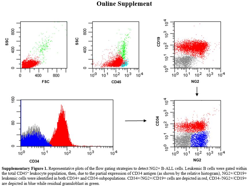
Only 2 pro-B ALLs with KMT2A-r showed low NG2 expression (<below the 10%) positivity cut-off, being equal to 4% and 5%, probably due to the steroid therapy administered to these patients before diagnostic work-up. The remaining 3/88 NG2+ B-ALLs (3.4%) did not harbor KMT2A-r– (1 pro-B and 2 B-common ALLs).
Antigen detection and surface expression intensity. To evaluate if the presence of KMT2A-r was associated with a distinctive immunophenotypic profile, beyond  NG2+ B-ALLs, we analyzed and compared the MFI of CD45, TdT, CD34, CD19, CD20, CD22, CD38, CD58 and, CD123 antigens in NG2+ KMT2A-r + (n=85; 77 pro-B and 8 B-common ALLs) and NG2– KMT2A-r – B-ALLs (n= 39; 12 pro-B and 27 B-common ALLs).  As shown in Table 1. In addition, to exclude any influence of the B-ALLs EGIL subtype on the peculiar immunophenotypic profile of KMT2Ar+ cases, we compared the expression intensity of all the markers analyzed, amongst the same subtype of B-ALLs (Table 2). Most of the markers analyzed (7/9, 77.8%) were differently expressed between the two categories. CD45 was expressed in all cases: MFI values were significantly higher in KMT2A-r+ than KMT2A-r– B-ALL regardless of the EGIL immunologic subtypes. CD34 was partially expressed in KMT2Ar+. More specifically, KMT2Ar+ B-ALLs were characterized by a significantly lower percentage of CD34+ blasts, as well as MFI, respect to KMT2Ar– samples independently from the EGIL subtype. In addition, among our cases, CD34 MFI appeared significantly lower in the presence of KMT2Ar, independently from the B-ALLs EGIL immunologic subtype (Table 2). TdT was expressed in all cases of B-lineage ALL. In particular, KMT2Ar+ B-ALL showed a significant downmodulation of TdT MFI compared to KMT2Ar– cases. This evidence was also confirmed within the same immunologic EGIL subtypes (KMT2Ar+ vs KMT2Ar– pro-B ALL) (Table 2).


Table 1 
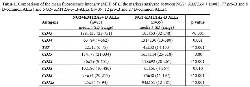
Table 1. Comparison of the mean fluorescence intensity (MFI) of all the markers analyzed between NG2+ KMT2A-r+ (n=85; 77 pro-B and 8 B-common ALLs) and NG2– KMT2A-r– B-ALLs (n= 39; 12 pro-B and 27 B-common ALLs).

Table 2 
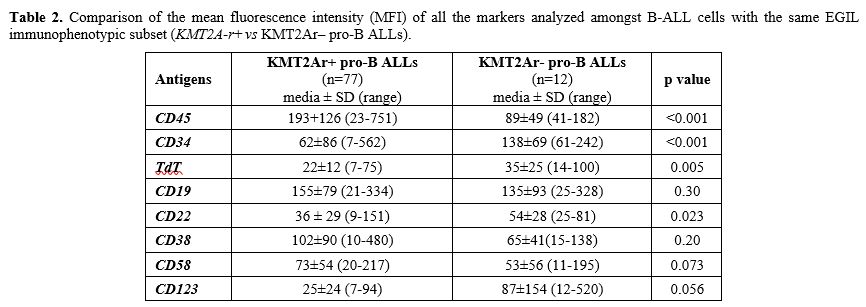
Table 2. Comparison of the mean fluorescence intensity (MFI) of all the markers analyzed amongst B-ALL cells with the same EGIL immunophenotypic subset (KMT2A-r+ vs KMT2Ar– pro-B ALLs).

CD19 and CD22, both targetable antigens, were expressed in all cases; interestingly CD19 was equally expressed in terms of both percentage and MFI regardless of KMT2A-r status; at variance, CD22 MFI values were significantly lower in KMT2Ar+ than KMT2Ar– B-ALLs, thus at least explaining the suboptimal efficacy of inotuzumab in this set of patients.[16]
CD38 was positive in all B-ALLs studied, and its MFI was significantly higher in (KMT2A-r+ vs KMT2A-r–: 102±90 vs 65±56, p=0.010). Finally, CD58 MFI appeared higher in KMT2Ar+ B-ALL (KMT2Ar+ vs KMT2Ar–: 73±54 vs 52±48, p< 0.001) while CD123, the interleukin-3 (IL-3) receptor α-chain, another antigen useful in MRD, had a lower MFI in KMT2Ar (KMT2Ar+ vs KMT2Ar–: 25±24 vs 94±151, p< 0.001).
Two additional markers were also evaluated: CD15 was evaluated  in 48/88 NG2+ cases (43 KMT2A::AFF1+ pro-B, 3 KMT2A::AFF1+ B-common and 2 KMT2A::MLLT1+ pro-B ALLs) and detected in in 16/48 NG2+ cases (CD15+ B-ALLs: 72.06±24.40%, range 26-90%; CD15 MFI: 78.16±53.4, range 20-190);  CD133 (Prominin-1), , was again studied in 48/88 NG2+ cases (43 KMT2A::AFF1+ pro-B, 3 KMT2A::AFF1+ B-common and 2 KMT2A::MLLT1+ pro-B ALLs) and was detected in all cases. with a mean percentage of positive cells of 84.37 ±11.64 (range 55-97%) and a mean MFI of 92.42±45.29 (range 18-190), suggesting a strong association of CD133 expression with NG2 positivity and the presence of KMT2Ar, mainly in the pro-B EGIL subtype.
Moreover, no differences in any of the antigens analysed were observed comparing KMT2Ar+ (n=8) vs KMT2Ar– (n=27) B-common ALLs, probably due to the low number of KMT2Ar+ cases characterized by this subtype (Table 3). In addition, no differences emerged in terms of antigen expression between KMT2Ar– pro-B ALLs and KMT2Ar– B-common ALLs, confirming the key role played by the presence of KMT2Ar in the peculiar phenotype of KMT2Ar+ leukemic cells (Table 4).


Table 3 
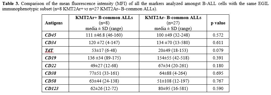
Table 3. Comparison of the mean fluorescence intensity (MFI) of all the markers analyzed amongst B-ALL cells with the same EGIL immunophenotypic subset (n=8 KMT2Ar+ vs n=27 KMT2Ar‒ B-common ALLs).
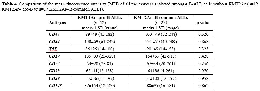
Table 4 Table 4.  Comparison of the mean fluorescence intensity (MFI) of all the markers analyzed amongst B-ALL cells without KMT2Ar (n=12 KMT2Ar‒ pro-B vs n=27 KMT2Ar– B-common ALLs).


Discussion

Immunophenotypic characterization of ALL is crucial for diagnosis and prognosis. This study identifies an immunophenotypic pattern that can be useful for the rapid identification of B-ALLs with a strong correlation between NG2 positivity and KMT2Ar in diagnostic routines. Other studies have demonstrated the high sensitivity of NG2 in identifying KMT2Ar+ B-ALLs. However, most of them were performed on relatively small samples or very heterogeneous groups.[17-18] Therefore, the strength of our study lies in a homogeneous and large cohort of newly diagnosed patients referred to a single Institution. NG2 belongs to the chondroitin sulfate proteoglycan family. It is highly expressed during early embryonic development but downregulated during differentiation. Its role in normal hematopoiesis and MLL-mediated leukemogenesis is still unclear.[19-21] Originally, NG2 was found to be highly expressed in melanoma patients and associated with tumor cell adhesion, migration, and metastasis.[13,22,23] In KMT2A-r+ B-ALLs, NG2 expression has recently been shown to be involved in leukemia invasiveness and central nervous system (CNS) infiltration, contributing to the frequent occurrence of CNS disease/relapse in this type of B-ALLs. The clinical data from the infant cohort of KMT2Ar+ B-ALL showed that high NG2 expression is associated with lower event-free survival, a higher number of circulating blasts and a more frequent occurrence of CNS disease/relapse. This is because NG2 expression is highly upregulated in blasts infiltrating extramedullar hematopoietic sites and CNS. In our study, 88/1382 (6.4%) cases displayed NG2 positivity with a cut-off more than 10%. As this marker is not uniformly expressed in the leukemic cell population, different levels of positivity may have predictive value. Our threshold level for NG2 positivity was in line with the work by Zerkalenkova E et al.,[24] in which the authors suggest that while any detectable NG2 positivity strongly associates with the presence of KMT2Ar in infant ALLs, NG2 positivity above 10% should be considered predictive of KMT2Ar in other ALL patient groups. Therefore, subsequent molecular and/or cytogenetic analysis should be performed in these patients.
Additionally, we also evaluated NG2 MFI. The percentage of NG2+ blasts is a more feasible and reliable predictor of KMT2Ar+ B-ALLs than NG2 expression quantification. Therefore, we considered NG2 positivity expressed as a percentage of positive cells.
In our cohort, 85/88 NG2+ cases were characterized by KMT2Ar+, while 3/88 cases, although NG2+, appeared KMT2Ar‒ by molecular analysis. However, we could not exclude the presence of KMT2Ar even in the latter cases, as cytogenetic data were not available. Furthermore, only two cases of B-ALL with NG2 expression below the 10% positivity threshold (4% and 5%, respectively), were found to be KMT2Ar+, likely due to the steroid therapy administration before diagnosis to these patients, which is known to cause down-modulation of this antigen.
The steroid-induced downmodulation together with its characteristic heterogeneous expression on the leukemic cell population, makes NG2 a reliable marker for the identification of KMT2Ar+ B-ALLs in therapy-naive patients at diagnosis; on the contrary, this antigen should not be considered for MRD evaluation. Accordingly, NG2 evaluation plays a crucial role into the diagnostic workflows for leukemia immunophenotyping, due to its strong positive predictive value for KMT2Ar, leading to the prompt identification of this subgroup of patients[25] who often need stem cells transplantation because of their dismal prognosis. In this subset of B-ALLs, NG2 appeared also associated with CD133 antigen expression.[26]
Based on our experience, the association between CD133 and NG2 is highly specific to KMT2Ar+ leukemic cells. This association strengthens the role of NG2 positivity in predicting KMT2Ar+ B-ALL. In addition to NG2 expression, our data also revealed that KMT2Ar+ differ from KMT2Ar‒ B-ALLs also in other phenotypic characteristics, such as the presence/absence of certain antigens and their varying expression intensity. Specifically, CD45, CD38, and CD58 show higher expression intensity in the presence of KMT2Ar, while CD34, CD22, TdT, and CD123 show lower expression intensity compared to KMT2Ar‒ cases. On the contrary, as previously described,[27] no differences were observed in CD19 MFI between the two categories. Therefore, as the presence of KMT2Ar increases CD38 MFI and does not impact the intensity of CD19 antigen expression, these patients can still benefit from the specific therapy currently available (monoclonal antibodies).[28-29] On the other hand, no differences in any of the antigens analysed were observed comparing KMT2Ar+ vs KMT2Ar– B-common ALLs, probably due to the low number of KMT2Ar+ cases characterized by this EGIL immunologic subtype. However, no differences were observed in any of the analysed antigens when comparing KMT2Ar+ and KMT2Ar- B-common ALLs. This is likely due to the low number of KMT2Ar+ cases characterised by this EGIL immunologic subtype. In addition, a recent study also shows the crucial role of NG2 in leukemogenesis and its potential role as specific antigen of a target therapy. Blockade of NG2 would seem to remove the chemoprotective effect of the bone marrow stroma through the mobilization of KMT2Ar+ blasts in the peripheral blood, which thus become more accessible to chemotherapy.[30] Overall, our data show the important role of NG2 detection since this antigen leads to a precise and reliable prediction of high-risk adult B-ALL patients characterized by KMT2Ar. In conclusion NG2 should be always included in the diagnostic MFC panel in combination with CD133 for the rapid detection of this peculiar and poor prognosis B-ALL subset.


Acknowledgments

MLM and SI were supported by ROMAIL ONLUS.

Author Contributions

Study concept and design: MSDP; flow cytometry assessment: MLM, SI, MSDP; data collection, acquisition, analysis, and interpretation: MLB, NP, MSDP; molecular data analysis and interpretation: LE; samples collection: MDT; molecular data collection and acquisition: M. Matarazzo; manuscript preparation: MLB, NP, MSDP; contribution to the lab work: FM; supervision and manuscript editing: M. Martelli. All authors have read and agreed to the published version of the manuscript.

References   

  1. Hoelzer D, Bassan R, Dombret H, Fielding A, Ribera JM, Buske C; ESMO Guidelines Committee. Acute lymphoblastic leukaemia in adult patients: ESMO Clinical Practice Guidelines for diagnosis, treatment and follow-up. Ann Oncol. 2016 Sep;27(suppl 5):v69-v82. https://doi.org/10.1093/annonc/mdw025 PMid:27056999
  2. Szczepański T, van der Velden VH, van Dongen JJ. Flow-cytometric immunophenotyping of normal and malignant lymphocytes. Clin Chem Lab Med. 2006;44(7):775-96. https://doi.org/10.1515/CCLM.2006.146 PMid:16776621
  3. Coustan-Smith E, Behm FG, Sanchez J, Boyett JM, Hancock ML, Raimondi SC, Rubnitz JE, Rivera GK, Sandlund JT, Pui CH, Campana D. Immunological detection of minimal residual disease in children with acute lymphoblastic leukaemia. Lancet. 1998 Feb 21;351(9102):550-4. https://doi.org/10.1016/S0140-6736(97)10295-1 PMid:9492773
  4. Kulis J, Sędek Ł, Słota Ł, Perkowski B, Szczepański T. Commonly Assessed Markers in Childhood BCP-ALL Diagnostic Panels and Their Association with Genetic Aberrations and Outcome Prediction. Genes (Basel). 2022 Jul 31;13(8):1374. https://doi.org/10.3390/genes13081374 PMid:36011285 PMCid:PMC9407579
  5. Richard-Carpentier G, Kantarjian HM, Tang G, Yin CC, Khoury JD, Issa GC, Haddad F, Jain N, Ravandi F, Short NJ, DiNardo CD, Takahashi K, Konopleva MY, Daver NG, Kadia T, Garcia-Manero G, Garris R, O'Brien S, Jabbour E. Outcomes of acute lymphoblastic leukemia with KMT2A (MLL) rearrangement: the MD Anderson experience. Blood Adv. 2021 Dec 14;5(23):5415-5419. https://doi.org/10.1182/bloodadvances.2021004580 PMid:34525185 PMCid:PMC9153023
  6. Attarbaschi A, Möricke A, Harrison CJ, Mann G, Baruchel A, De Moerloose B, Conter V, Devidas M, Elitzur S, Escherich G, Hunger SP, Horibe K, Manabe A, Loh ML, Pieters R, Schmiegelow K, Silverman LB, Stary J, Vora A, Pui CH, Schrappe M, Zimmermann M; Childhood Acute Lymphoblastic Leukemia Working Group. Outcomes of Childhood Noninfant Acute Lymphoblastic Leukemia With 11q23/KMT2A Rearrangements in a Modern Therapy Era: A Retrospective International Study. J Clin Oncol. 2023 Mar 1;41(7):1404-1422. https://doi.org/10.1200/JCO.22.01297 PMid:36256911
  7. Piciocchi A, Messina M, Elia L, Vitale A, Soddu S, Testi AM, Chiaretti S, Mancini M, Albano F, Spadano A, Krampera M, Bonifacio M, Cairoli R, Vetro C, Colella F, Ferrara F, Cimino G, Bassan R, Fazi P, Vignetti M. Prognostic impact of KMT2A-AFF1-positivity in 926 BCR-ABL1-negative B-lineage acute lymphoblastic leukemia patients treated in GIMEMA clinical trials since 1996. Am J Hematol. 2021 Sep 1;96(9):E334-E338. https://doi.org/10.1002/ajh.26253
  8. Motlló C, Ribera JM, Morgades M, Granada I, Montesinos P, Brunet S, Bergua J, Tormo M, García-Boyero R, Sarrà J, Del Potro E, Grande C, Barba P, Bernal T, Amigo ML, Grau J, Cervera J, Feliu E; PETHEMA Group, Spanish Society of Hematology. Frequency and prognostic significance of t(v;11q23)/KMT2A rearrangements in adult patients with acute lymphoblastic leukemia treated with risk-adapted protocols. Leuk Lymphoma. 2017 Jan;58(1):145-152. https://doi.org/10.1080/10428194.2016.1177182 PMid:27122129
  9. Meyer C, Larghero P, Almeida Lopes B, Burmeister T, Gröger D, Sutton R , Venn N.C., Cazzaniga G, Corral Abascal L,Tsaur G, Fechina L, Emerenciano M, Pombo-de-Oliveira, M.S, Lund-Aho T, Lundán T, Montonen M, Juvonen V, Zuna J, Trka J, Ballerini P, Lapillonne H, Van der Velden VHJ, Sonneveld E, Delabesse E,de Matos RRC, Silva MLM, Bomken S, Katsibardi K, Keernik M, Grardel N,Mason J,Price R, Kim J, Eckert C, Lo Nigro L,Bueno C,Menendez P,zur Stadt U, Gameiro P, Sedék L, Szczepański T, Bidet A, Marcu V, Shichrur K, Izraeli S, Madsen HO, Schäfer BW, Kubetzko S, Kim R, Clappier E, Trautmann H, Brüggemann M, Archer P, Hancock J, Alten J, Möricke A, Stanulla M, Lentes J, Bergmann AK, Strehl S, Köhrer S, Nebral K,Dworzak M.N, Haas OA, Arfeuille C, Caye-Eude A, Cavé H, Marschalek R. The KMT2A recombinome of acute leukemias in 2023.Leukemia. 2023; 37(5): 988-1005. https://doi.org/10.1038/s41375-023-01877-1 PMid:37019990 PMCid:PMC10169636
  10. Zangrando, A., Dell'Orto, M.C., te Kronnie, G., Basso G. MLL rearrangements in pediatric acute lymphoblastic and myeloblastic leukemias: MLL specific and lineage specific signatures. BMC Med Genomics. 2009 Jun 23;2:36. https://doi.org/10.1186/1755-8794-2-36 PMid:19549311 PMCid:PMC2709660
  11. Pui, C.H.; Frankel, L.S.; Carroll, A.J.; Raimondi, S.C.; Shuster, J.J.; Head, D.R.; Crist, W.M.; Land, V.J.; Pullen, D.J.; Steuber, C.P. Clinical characteristics and treatment outcome of childhood acute lymphoblastic leukemia with the t(4;11)(q21;q23): A collaborative study of 40 cases. Blood 1991, 77, 440-447. https://doi.org/10.1182/blood.V77.3.440.440 PMid:1991161
  12. Smith FO, Rauch C, Williams DE, March CJ, Arthur D, Hilden J, Lampkin BC, Buckley JD, Buckley CV, Woods WG, Dinndorf PA, Sorensen P, Kersey J, Hammond D, Bernstein ID. The human homolog of rat NG2, a chondroitin sulfate proteoglycan, is not expressed on the cell surface of normal hematopoietic cells but is expressed by acute myeloid leukemia blasts from poor-prognosis patients with abnormalities of chromosome band 11q23. Blood. 1996 Feb 1;87(3):1123-33. https://doi.org/10.1182/blood.V87.3.1123.bloodjournal8731123 PMid:8562938
  13. Behm FG, Smith FO, Raimondi SC, Pui CH, Bernstein ID. The human homolog of the rat chondroitin sulfate proteoglycan, NG2, detected by monoclonal antibody 7.1, identifies childhood acute lymphoblastic leukemias with t(4;11)(q21;q23) or t(11;19)(q23;p13) and MLL gene rearrangements. Blood. 1996 Feb 1;87(3):1134-9. https://doi.org/10.1182/blood.V87.3.1134.bloodjournal8731134 PMid:8562939
  14. Prieto C, López-Millán B, Roca-Ho H, Stam RW, Romero-Moya D, Rodríguez-Baena FJ, Sanjuan-Pla A, Ayllón V, Ramírez M, Bardini M, De Lorenzo P, Valsecchi MG, Stanulla M, Iglesias M, Ballerini P, Carcaboso ÁM, Mora J, Locatelli F, Bertaina A, Padilla L, Rodríguez-Manzaneque JC, Bueno C, Menéndez P. NG2 antigen is involved in leukemia invasiveness and central nervous system infiltration in MLL-rearranged infant B-ALL. Leukemia. 2018 Mar;32(3):633-644. doi: 10.1038/leu.2017.294. Epub 2017 Sep 25. Erratum in: Leukemia. 2018 Oct;32(10):2306. https://doi.org/10.1038/leu.2017.294 PMid:28943635 PMCid:PMC5843903
  15. Elia, L., Mancini, M., Moleti, L., Meloni, G., Buffolino, S., Krampera, M., De Rossi, G., Foà, R. & Cimino, G. (2003) A multiplex reverse transcriptase-polymerase chain reaction strategy for the diagnostic molecular screening of chimeric genes: a clinical evaluation on 170 patients with acute lymphoblastic leukemia. Haematologica, 88, 275-279.
  16. Kantarjian HM, DeAngelo DJ, Stelljes M, Martinelli G, Liedtke M, Stock W, Gökbuget N, O'Brien S, Wang K, Wang T, Paccagnella ML, Sleight B, Vandendries E, Advani AS. Inotuzumab Ozogamicin versus Standard Therapy for Acute Lymphoblastic Leukemia. N Engl J Med. 2016 Aug 25;375(8):740-53. https://doi.org/10.1056/NEJMoa1509277 PMid:27292104 PMCid:PMC5594743
  17. Zangrando A, Intini F, te Kronnie G, Basso G. Validation of NG2 antigen in identifying BP-ALL patients with MLL rearrangements using qualitative and quantitative flow cytometry: a prospective study. Leukemia. 2008; 22: 858-61. https://doi.org/10.1038/sj.leu.2404952 PMid:17851550
  18. Emerenciano, M.; Renaud, G.; Sant'Ana, M.; Barbieri, C.; Passetti, F.; Pombo-de-Oliveira, M.S. Challenges in the Use of NG2 Antigen as a Marker to Predict MLL Rearrangements in Multi-Center Studies. Leuk. Res. 2011, 35, 1001-1007. https://doi.org/10.1016/j.leukres.2011.03.006 PMid:21444110
  19. Schwartz, S.; Rieder, H.; Schläger, B.; Burmeister, T.; Fischer, L.; Thiel, E. Expression of the Human Homologue of Rat NG2 in Adult Acute Lymphoblastic Leukemia: Close Association with MLL rearrangement and a CD10(-)/CD24(-)/CD65s(+)/CD15(+) B-cell phenotype. Leukemia. 2003 Aug;17(8):1589-95. https://doi.org/10.1038/sj.leu.2402989 PMid:12886247
  20. Thirman MJ, Gill HJ, Burnett RC, Mbangkollo D, McCabe NR, Kobayashi H, Ziemin-van der Poel S, Kaneko Y, Morgan R, Sandberg AA, Chaganti RSK, Larson RA, Le Beau MM, Diaz MO, Rowley ID: Rearrangements of the MLL gene in acute lymphoblastic and acute myeloid leukemias with 1lq23 chromosomal translocations. N Engl J Med 329:909, 1993. https://doi.org/10.1056/NEJM199309233291302 PMid:8361504
  21. Bueno C, Montes R, Martín L, Prat I, Hernandez MC, Orfao A, Menendez P. NG2 antigen is expressed in CD34+ HPCs and plasmacytoid dendritic cell precursors: is NG2 expression in leukemia dependent on the target cell where leukemogenesis is triggered? Leukemia. 2008 Aug;22(8):1475-8. https://doi.org/10.1038/leu.2008.134 PMid:18698324
  22. Menendez P, Bueno C. Expression of NG2 antigen in MLL-rearranged acute leukemias: how complex does it get? Leuk Res 2011; 35: 989-990. https://doi.org/10.1016/j.leukres.2011.03.015 PMid:21492935
  23. Price MA, Colvin Wanshura LE, Yang J, Carlson J, Xiang B, Li G et al. CSPG4, a potential therapeutic target, facilitates malignant progression of melanoma. Pigment Cell Melanoma Res 2011; 24: 1148-1157. https://doi.org/10.1111/j.1755-148X.2011.00929.x PMid:22004131 PMCid:PMC3426219
  24. Zerkalenkova E, Mikhaylova E, Lebedeva S, Illarionova O, Baidun L, Kashpor S, Osipova E, Maschan M, Maschan A, Novichkova G, Olshanskaya Y, Popov A. Quantification of NG2-positivity for the precise prediction of KMT2A gene rearrangements in childhood acute leukemia. Genes Chromosomes Cancer. 2021; 60(2):88-99. https://doi.org/10.1002/gcc.22915 PMid:33135273
  25. Tsagarakis NJ, Papadhimitriou SI, Pavlidis D, Marinakis T, Kostopoulos IV, Stiakaki E, Polychronopoulou S, Paterakis G. Flow cytometric predictive scoring systems for common fusions ETV6/RUNX1, BCR/ABL1, TCF3/PBX1 and rearrangements of the KMT2A gene, proposed for the initial cytogenetic approach in cases of B-acute lymphoblastic leukemia. Int J Lab Hematol. 2019 Jun;41(3):364-372. https://doi.org/10.1111/ijlh.12983 PMid:30730614
  26. Godfrey L, Crump NT, O'Byrne S, Lau IJ, Rice S, Harman JR, Jackson T, Elliott N, Buck G, Connor C, Thorne R, Knapp DJHF, Heidenreich O, Vyas P, Menendez P, Inglott S, Ancliff P, Geng H, Roberts I, Roy A, Milne TA. H3K79me2/3 controls enhancer-promoter interactions and activation of the pan-cancer stem cell marker PROM1/CD133 in MLL-AF4 leukemia cells. Leukemia. 2021 Jan;35(1):90-106. https://doi.org/10.1038/s41375-020-0808-y PMid:32242051 PMCid:PMC7787973
  27. Raponi S, De Propris MS, Intoppa S, Milani ML, Vitale A, Elia L, Perbellini O, Pizzolo G, Foá R, Guarini A. Flow cytometric study of potential target antigens (CD19, CD20, CD22, CD33) for antibody-based immunotherapy in acute lymphoblastic leukemia: analysis of 552 cases. Leuk Lymphoma. 2011 Jun;52(6):1098-107. https://doi.org/10.3109/10428194.2011.559668 PMid:21348573
  28. Voruz S, Blum S, de Leval L, Schoumans J, Solly F, Spertini O. Daratumumab and venetoclax in combination with chemotherapy provide sustained molecular remission in relapsed/refractory CD19, CD20, and CD22 negative acute B lymphoblastic leukemia with KMT2A-AFF1 transcript. Biomark Res. 2021 Dec 20;9(1):92. https://doi.org/10.1186/s40364-021-00343-3 PMid:34930453 PMCid:PMC8686620
  29. Boissel N, Chiaretti S, Papayannidis C, Ribera JM, Bassan R, Sokolov AN, Alam N, Brescianini A, Pezzani I, Kreuzbauer G, Zugmaier G, Foà R, Rambaldi A. Real-world use of blinatumomab in adult patients with B-cell acute lymphoblastic leukemia in clinical practice: results from the NEUF study. Blood Cancer J. 2023 Jan 4;13(1):2. https://doi.org/10.1038/s41408-022-00766-7 PMid:36599847 PMCid:PMC9813344
  30. Lopez-Millan B, Sanchéz-Martínez D, Roca-Ho H, Gutiérrez-Agüera F, Molina O, Diaz de la Guardia R, Torres-Ruiz R, Fuster JL, Ballerini P, Suessbier U, Nombela-Arrieta C, Bueno C, Menéndez P. NG2 antigen is a therapeutic target for MLL-rearranged B-cell acute lymphoblastic leukemia. Leukemia. 2019; 33(7):1557-1569 https://doi.org/10.1038/s41375-018-0353-0 PMid:30635633 PMCid:PMC6755967

Supplementary Files


Suppl Fig 1
Supplementary Figure 1. Representative plots of the flow gating strategies to detect NG2+ B-ALL cells. Leukemic B cells were gated within the total CD45+ leukocyte population, then, due to the partial expression of CD34 antigen (as shown by the relative histogram), NG2+/CD19+ leukemic cells were identified in both CD34+ and CD34-subpopulations. CD34+/NG2+/CD19+ cells are depicted in red, CD34-/NG2+/CD19+ are depicted in blue while residual granuloblast in green.

In the remaining 23 patients (22 males and 1 female, years range 20-82), representing 19.45% of the total cohort, more than