Coexistence of Multiple Gene Variants in Some Patients with Erythrocytoses

Andrea Benetti1, Irene Bertozzi1, Giulio Ceolotto2, Irene Cortella1, Daniela Regazzo1, Giacomo Biagetti1, Elisabetta Cosi1 and Maria Luigia Randi1.

1 First Medical Clinic, Department of Medicine – DIMED, University of Padova, Padova, Italy
2 Emergency Medicine, Department of Medicine – DIMED, University of Padova, Padova, Italy.
 

Correspondence to: Maria Luigia Randi, Department of Medicine (DIMED), via Giustiniani 2, 35128 Padova, Italy. Tel: +39 3493986088. E-mail: marialuigia.randi@unipd.it

Published: March 01, 2024
Received: November 03, 2023
Accepted: February 10, 2024
Mediterr J Hematol Infect Dis 2024, 16(1): e2024021 DOI 10.4084/MJHID.2024.021

This is an Open Access article distributed under the terms of the Creative Commons Attribution License
(
https://creativecommons.org/licenses/by-nc/4.0), which permits unrestricted use, distribution, and reproduction in any medium, provided the original work is properly cited.

Abstract

Background: Erythrocytosis is a relatively common condition; however, a large proportion of these patients (70%) remain without a clear etiologic explanation. 
Methods:
We set up a targeted NGS panel for patients with erythrocytosis, and 118 sporadic patients with idiopathic erythrocytosis were studied.
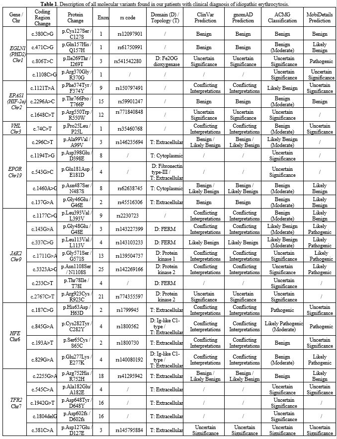
Results:
In 40 (34%) patients, no variant was found, while in 78 (66%), we identified at least one germinal variant; 55 patients (70.5%) had 1 altered gene, 18 (23%) had 2 alterations, and 5 (6.4%) had 3. An 
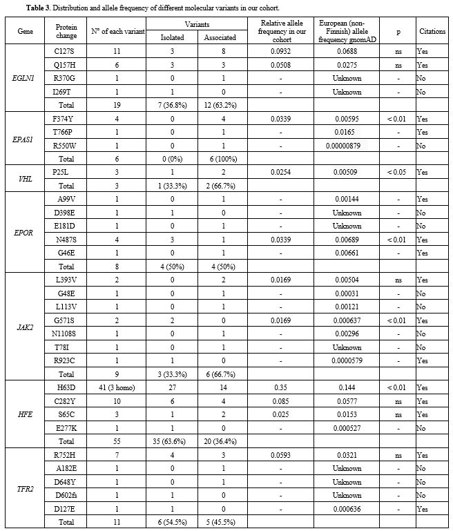
altered HFE gene was observed in 51 cases (57.1%), EGLN1 in 18 (22.6%) and EPAS1, EPOR, JAK2, and TFR2 variants in 7.7%, 10.3%, 11.5%, and 
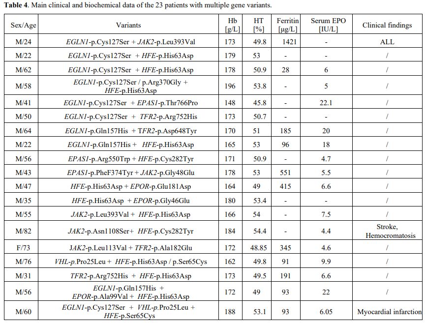
14.1% patients, respectively. In 23 patients (19.45%), more than 1 putative variant was found in multiple genes.
Conclusions:
Genetic variants in patients with erythrocytosis were detected in about 2/3 of our cohort. An NGS panel including more candidate genes should reduce the number of cases diagnosed as “idiopathic” erythrocytosis in which a cause cannot yet be identified. 
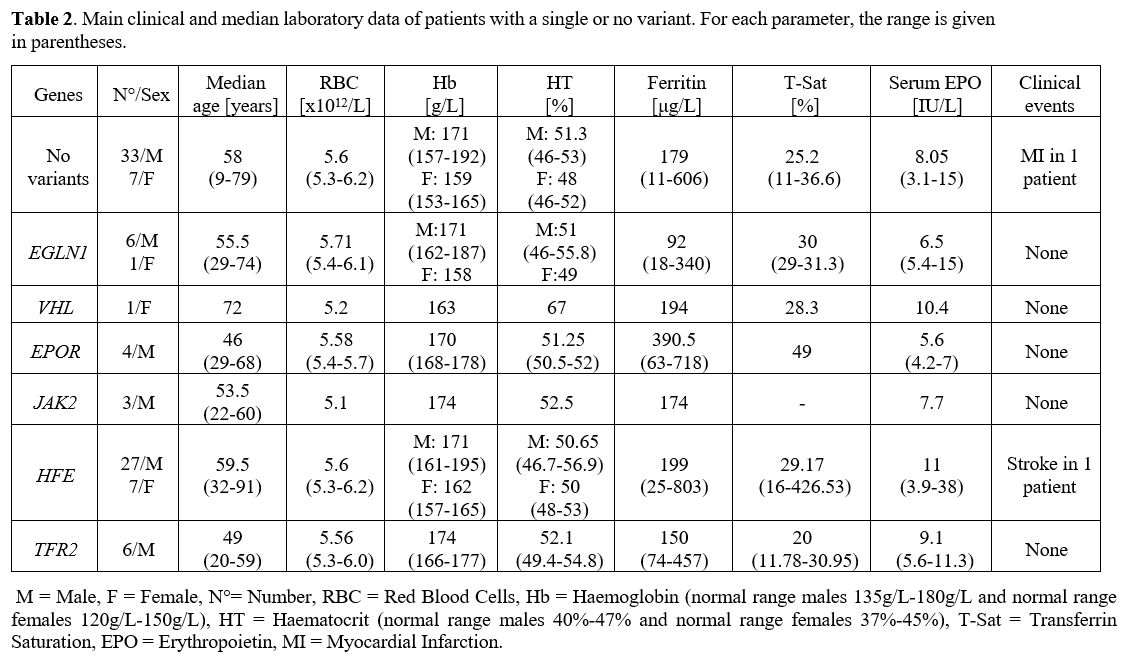
It is known that HFE variants are common in idiopathic erythrocytosis. TFR2 alterations support the existence of a relationship between genes involved in iron metabolism and impaired erythropoiesis. Some novel multiple variants were identified. Erythrocytosis appears to be often multigenic.


Introduction

Erythrocytosis, characterised by increased haemoglobin (Hb) and/or haematocrit (HT) levels, is a relatively common condition in the general population;[1] some of these patients are affected by polycythaemia vera (PV);[2] acquired secondary erythrocytosis are more frequent and are due to increased levels of erythropoietin (EPO) appropriately or inappropriately produced. Finally, congenital erythrocytosis may be the consequence of mutations of the globin genes (HBB or HBA), or bi-phosphoglycerate mutase (BPGM),[3] or of gene mutations involved in the oxygen sensing pathway (VHL, PHD2, HIF2A) or of the erythropoietin receptor gene (EPOR).[4]
Unfortunately, a large proportion (70%) of patients with erythrocytosis remains without a clear aetiologic explanation[5] and, consequently, the condition has been classified as idiopathic erythrocytosis (IE).[6]
We observed that a significant percentage of IE patients have high or borderline high ferritin levels, suggesting that at least in some of the patients, erythrocytosis coexists with an impaired iron balance, and HFE gene mutations are higher in IE than in the general population.[7]
The availability of next-generation sequencing (NGS) facilities in our laboratory prompted us to build a specific gene panel to search for novel variants in patients with erythrocytosis of uncertain aetiology.[8–10]


Material and Methods

From 2013 to 2020, in our centre, we encountered 118 (M/F =101/17; mean age 53.7±17.2 years) patients who were diagnosed with idiopathic erythrocytosis. Patients’ inclusion criteria were: (i) haemoglobin >180 g/L and haematocrit  >52% in males and haemoglobin  >160 g/L and haematocrit  >46% in females, (ii) long-standing unexplained erythrocytosis, (iii) absence of relatives with increased Hb or HT, (iv) no evidence of smoke, arterial-venous shunt, pulmonary and/or renal diseases or neoplasms, (v) absence of somatic mutations in JAK2 (p.Val617Phe or variants in JAK2 exon 12). Patients carrying haemoglobin variants with high oxygen affinity, as evaluated by venous p50[1] and those with a left-shifted oxygen dissociation curve[11] were excluded. No children younger than 16 years of age are included in the present cohort, as our surgery is dedicated only to adults. We evaluated nine normal subjects (normal Hb/HT values and no clinical signs of erythrocytosis) as controls.
The patients' data were collected in an ad hoc performed anonymous database approved by the Ethics Committee (EC) of the General Hospital-University of Padova, Italy. The patients gave consent to data collection for scientific purposes and publication. The study was conducted in accordance with the Declaration of Helsinki.
Thrombotic and haemorrhagic events were recorded. We collected red blood cell (RBC) counts, Hb and HT levels in all patients, ferritin levels in 67, transferrin saturation (T-Sat) in 38, and serum erythropoietin (EPO) in 66.
According to the manufacturer's instructions, DNA was extracted from granulocytes obtained from patients’ blood samples using EuroGold Blood DNA Mini Kit Plus (EuroClone) and stored at -20°C. DNA concentration was determined by Qubit 4 Fluorometer with QubitTM 1X dsDNA HS Assay Kits (Thermo Fisher Scientific).
A targeted NGS panel for patients with erythrocytosis was set up. The panel included the coding sequence of 14 genes (BPGM, EGLN1, EPAS1, HAMP, FTL, HFE, HFE2, JAK2, SLC11A2, SLC40A1, TFR2, VHL, FTH1, EPOR) involved in erythrocytosis because we choose to obtain a targeted sequence of genes interested in a specific group of patients. The panel is an "On Demand AmpliSeq Panel" designed with the Illumina Design Studio platform and validated in silico by Illumina. Libraries were prepared by using "AmpliSeq for Illumina On-Demand, Custom, and Community Panels" (Illumina) as the manufacturer's instructions. The sequencing was performed in a MiSeq Illumina instrument (151 cycles paired-end; mean target coverage 500X for each sample). FASTAQ data were analysed by Illumina BaseSpace softwares and by CLC Genomics Workbench Qiagen tools. Homo sapiens genome assembly GRCh37 (hg19) was used as a reference, and Integrative Genomics Viewer (IGV) was used to visualise reads alignment. Germline and somatic variants were filtered using two independent and different workflows primarily based on different frequency thresholds for variant identification. In both workflows, the selection of variants was carried out through multiple filters that mainly consider the total coverage of the fragment of interest, the quality and the correct and minimum number of base calls. Only non-synonymous variants that passed all filters were considered for further validation and analysis. Germline variants identified by NGS were confirmed by direct Sanger sequencing.
Amplification conditions and primers are available upon request. The clinical significance of the identified variants was inferred using ClinVar, the OMIM database, MobiDetails, and gnomAD, according to ACMG guidelines[12] with the help of VARSOME.[13] The statistical analysis was performed by comparing the data obtained in patients and controls with a Mann-Whitney U test (two groups).


Results

At the time of the study, the patients’ median Hb was 172 g/L (range 142-206) and HT 51% (range 45.7-57.2). While in 40 (34%) patients, we did not detect any gene variant, in 78 (66%), we identified at least one germinal variant in one of the studied genes (Table 1). Out of the 78 patients with germinal variants, 55 (70.5%) had 1 altered gene, 18 (23.1%) had 2 alterations, and 5 (6.4%) had 3. We did not find significant differences in the laboratory data and clinical characteristics between patients with or without alterations, and the number of variants had no impact on patients' parameters. The main clinical and laboratory data of patients with an undetected variant and patients with a single variant are summarised in Table 2

Table 1 Table 1. Description of all molecular variants found in our patients with clinical diagnosis of idiopathic erythrocytosis.

Table 2 Table 2. Main clinical and median laboratory data of patients with a single or no variant. For each parameter, the range is given in parentheses.

The most common altered gene in our cohort was HFE, present in 51 cases (65.3%) and, in particular, the presence of HFE-p.His63Asp was found in 41 (52.5% of all patients carrying variants). The altered EGLN1 gene was found in 18 (23.1%) patients, where p.Cys127Ser was the prevalent one (11 cases =61.1%). EPAS1, EPOR, JAK2, and TFR2 variants were observed in 6 (7.7%), 8 (10.3%), 9 (11.5%), and 11 (14.1%) patients, respectively (Table 3). 

Table 3
Table 3. Distribution and allele frequency of different molecular variants in our cohort.

In the remaining 23 patients (22 males and 1 female, years range 20-82), representing 19.45% of the total cohort, more than 1 putative variant was found in multiple genes (Table 4). 
In the control group, 7 subjects (78%) carry gene variants (4 EGLN1, 2 EPAS1-p.Thr766Pro, 2 JAK2-p.Leu393Val, 1 EPOR-p.Gly46Glu). Remarkably, no normal case displayed any HFE or TFR2 variant.


Table 4
Table 4. Main clinical and biochemical data of the 23 patients with multiple gene variants.


Discussion

In about 70% of patients with erythrocytosis, a specific aetiology remains elusive despite extensive testing.[14] Aberrant function of proteins involved in the oxygen-sensing pathway (PHD2, HIF2A, VHL, EPO), in EPOR gene, and rarely in PIEZO1 and SLC genes have been found in patients with sporadic erythrocytosis.[5] Thanks to the availability of the NGS technique, we studied 118 sporadic patients with a clinical diagnosis of IE. In our cohort, however, we found a high prevalence (>80%) of patients with altered genes known or supposed to be involved in familial erythrocytosis. Camps et al.[15] found erythrocytosis-causing variants in about half of their 125 patients and some novel variants in erythrocytosis-associated genes (LNK) using a targeted sequencing panel that did not include genes involved in iron metabolism. The higher percentage of patients with genetic alterations in our cohort is attributable to the common finding of iron metabolism gene variants; without their contribution, the frequency of variants is like Camps’ data.[15] The relevant number of patients with IE carrying HFE variants, whose mechanism remains to be elucidated, suggests that a relationship between molecules involved in iron regulation and erythrocytosis appears reasonably conceivable.
Oxygen sensing pathway genes. EGLN1 variants were found in 18 patients; more than half of them carried the p.Cys127Ser variant. Such a variant is common in Tibetans, often associated with variant D4E on the same allele, as it protects them against chronic mountain sickness, characterised by erythrocytosis, hypoventilation, and pulmonary hypertension.[16] The global frequency of this variant in our cohort is 0.0932, not different than that found in the general population (gnomAD) at normoxic conditions and in low-landers; the EGLN1-p.Cys127Ser variant is present in 15–30% of non-Tibetan controls, with a still unclear function[17] not associated with variant D4E as in our patients (https://www.ncbi.nlm.nih.gov/snp/rs12097901#frequency_tab). Six out of the 19 patients with EGLN1 variants (31,5%) carried EGLN1-p.Gln157His. This variant is mostly considered benign but described in 2 members of a family with MPN in association with JAK2-p.Val617Phe suggests that it should represent a disease-initiating event.[18] Neither germinal nor somatic-associated JAK2 variants were observed in our patients. In contrast, in 3 patients the EGLN1-p.Gln157His variant was associated with TFR2, HFE or EPOR alterations. In 2 patients we observed EGLN1-p.Arg370Gly and p.Ile269Thr variants, the first considered of unknown significance and the second as pathogenic using the MobiDetails annotation platform.[19]
A large number of EPAS1 variants have been observed and considered often benign or of uncertain significance. Some mosaic or somatic EPAS1 variants are associated with pheochromocytoma and/or paraganglioma (PPGL),[20] while gain-of-function alterations in exon 12 of the gene are known to cause familial erythrocytosis type 4 (ECYT4).[21] One of our patients presented the p.Arg550Trp variant, which has never been previously described; interestingly, a recent article[22] identified EPAS1-p.Arg550Gln variant in a patient with erythrocytosis and, considering that the two variants are in the same location, they interest a critical part of the protein and are in proximity to the pathogenetic variant identified by Percy et al.,[21] we speculate that they might be clinically significant. Four patients carried HIF2A-p.Phe374Tyr variant disrupting interaction with the Von Hippel–Lindau tumor suppressor (pVHL) in normoxian condition and 1 patient carried p.Thr766Pro already described in familial erythrocytosis but considered benign. Interestingly, EPAS1-p.Phe374Tyr and partially also EPAS1-p.Thr766Pro were stable in normoxia and retained their interaction with the pVHL.[20] EPAS1-p.Phe374Tyr variant seems particularly frequent in our erythrocytotic cohort: the number of cases is small and the comparison with general population loss it significance. Larger studies are needed to confirm this data.
It has been shown that the PHD2 mutation in Tibetan highlanders, in combination with EPAS1 polymorphism, lowers haemoglobin level concentration.[23] The authors suggested that, in Tibetan highlanders, as well as protected from polycythaemia, this genetic condition may confer physiological advantages, and the protection from erythrocytosis could be the reflection of ameliorated HIF-regulated activities.
Three sporadic patients had the VHL-p.Pro25Leu variant,[24] which is neither associated with Von Hipple Lindau syndrome nor with erythrocytosis. In 2 of our 3 cases with VHL-p.Pro25Leu the association with HFE-p.Ser65Cys was also highlighted, and in one, the association with EGLN1-p.Cys127Ser was also found.
EPOR gene. Four of our patients carried EPOR-p.Asn487Ser, which was already identified in familial erythrocytosis,[4,25] and is considered to play an important role in EPO signalling. The remaining four patients with EPOR variants had each p.Gly46Glu and p.Ala99Val (both considered benign) and p.Glu181Asp and p.Asp398Glu (previously undescribed). Half of the patients with alterations in EPOR presented additional variants in the genes of the panel. Of note, 4 patients with identified variants in EPOR also carried the HFE-p.His63Asp alteration.
JAK2 gene. While JAK2 somatic mutations are well-known driver mutations in Polycythaemia Vera (PV) and MPN,[26–29] the significance of germinal modifications of JAK2 remains unclear, as is the case for p.Gly48Glu (who also carried EPAS1-p.Phe374Tyr) and p.Asn1108Ser aminoacidic changes found in two of our patients. The p.Leu393Val variant, found in 2 patients, has been previously described in 1 patient who had documented normal blood counts for decades prior to his diagnosis of PV.[30] The authors hypothesised that the p.Leu393Val variant may precede the acquisition of JAK2-p.Val617Phe. However, the functional significance of this germline JAK2 variant and its putative predisposing role to PV remain unclear, though its potential to predispose to malignancy is suspected, as the germline JAK2 variant is frequently found in diffuse large B-cell tumours. Relying on these data, these patients are being closely followed up so that the appearance of PV can be promptly recognised. In addition to the somatic JAK2-p.Val617Phe mutation, other germline mutations can induce MPN phenotype or isolated thrombocytosis and erythrocytosis;[30] none of such germline variants have been found in our cohort and, at present, none of our patients displayed the JAK2-p.Val617Phe genotype. Some novel yet undescribed germinal variants of JAK2 were found in two cases associated with TFR2-p.Ala182Glu and with compound HFE variant plus heterozygous TFR2-p.Arg752His alteration.
Iron metabolism genes. HFE-p.His63Asp, present in two-thirds of our patients,[31,32] occurs with a significantly higher frequency than in the general population.[7] HFE-p.His63Asp variant is not capable of influencing the normal activity of hepcidin, the negative regulator of iron homeostasis,[33,34] but in patients with increased haematocrit levels, the variant could have a pathological effect. Fourteen patients with the HFE-p.His63Asp variant present also other gene variants, such as EGLN1, EPAS1, EPOR, JAK2, VHL and TFR2.
HFE-p.Cys282Tyr, p.Ser65Cys and p.Glu277Lys, both common in haemochromatosis,[33] were found in fewer patients. Three cases with compound HFE (2 p.Cys282Tyr/ p.His63Asp and 1 p.Ser65Cys/p.His63Asp) were variously associated with EPAS1 EGLN1, VHL, JAK2, TFR2. Of note, none of these cases had high serum ferritin, but all had a high haematocrit.[7] It is not clear whether the HFE variants represent a trigger sufficient to induce erythrocytosis, as suggested by the cases with single gene mutation and iron overload, as evidenced by increased ferritin. However, HFE variants are frequently observed in idiopathic erythrocytosis, and we speculate that they may somehow facilitate the increase in red blood cell mass.[7,31,35] Likewise, it is not clear whether the concomitance of iron metabolism gene variants with other gene mutations may represent a fertile ground for a putative effect on erythrocytosis.[36] Considering that iron is a critical component in the process of erythrocyte production, it may have a connection with erythrocytosis.[37] Indeed, relations between HAMP, EPAS1 and EGLN1 genes to control iron metabolism have already been observed in duodenal enterocytes.[38] Studies in other tissues will define the connection of these genes with increased erythropoiesis.
Several patients carried TFR2 alterations, the most frequent being p.Arg752His, which is considered benign and rarer in general European populations, though at present, little is known. Our finding needs to be confirmed in other cohorts of erythrocytotic patients. We found other 3 unknown variants. This gene resulted in both single and associated variants on other genes (EGLN1, JAK2, HFE). TFR2 has been identified as a component of the EPOR complex and is required for efficient erythropoiesis.[39] In mice, the loss of TFR2 in the erythroid compartment accounts for increased haemoglobin, indicating a deregulated erythropoiesis: it seems that TFR2 modulates the sensitivity of erythroid cells to erythropoietin (EPO) and endogenous EPO.[40,41] To the best of our knowledge, this is the first time that a mutated TFR2 has been found in patients with erythrocytosis, suggesting that, in some cases, TFR2 gene variants may impair erythropoiesis. Remarkably, TFR2 variants, not found in any of the normal cases, as HFE alterations, bring additional support to the potential relationship between genes involved in iron metabolism and erythrocytosis. Further studies are required to confirm this hypothesis.
Unfortunately, the available number of subjects without erythrocytosis evaluated with our NGS panel is small and inadequate to be statistically compared to our large cohort of erythrocytotic patients.
In the present study, the number of patients with variants is relatively high. However, among the total number of variants detected, 2 have a potentially pathogenic effect, and 5 are considered likely pathogenetic at least in one prediction tool.
Moreover, the gene alterations found do not have a clear link to erythrocytosis. These variants need to be further investigated to evaluate their functional effect.
The gene panel we used comprehends the coding region of each gene. However, targeted genic sequencing favoured a smaller set of data that was easier to evaluate, and we are now planning to perform whole genome sequencing in our patients.

Conclusions

The use of targeted NGS panels demonstrated that several diseases are characterised by multigenic mutations: cancers[42–44] dyslipidemia,[45] hystiocytosis,[46] etc. Our study shows that the occurrence of unexpected multiple variants, some of which are unreported yet, is common in patients with sporadic erythrocytosis. The exact role played by these variants in erythrocytosis remains to be further elucidated.

Acknowledgments

We acknowledge Dr. Agostino Faggiotto for his comments and contribution to the editing of the paper.

References   

  1. Rumi E, Passamonti F, Pagano L, et al. Blood p50 evaluation enhances diagnostic definition of isolated erythrocytosis. J Intern Med. 2009;265(2):266-274. https://doi.org/10.1111/j.1365-2796.2008.02014.x PMid:18793248
  2. Arber DA, Orazi A, Hasserjian R, et al. The 2016 revision to the World Health Organization classification of myeloid neoplasms and acute leukemia. Blood. 2016;127(20):2391-2405. https://doi.org/10.1182/blood-2016-03-643544 PMid:27069254
  3. Bento C, Percy MJ, Gardie B, et al. Genetic Basis of Congenital Erythrocytosis: Mutation Update and Online Databases. Hum Mutat. 2014;35(1):15-26. https://doi.org/10.1002/humu.22448 PMid:24115288
  4. Bento C. Genetic basis of congenital erythrocytosis. Int J Lab Hematol. 2018;40(January):62-67. https://doi.org/10.1111/ijlh.12828 PMid:29741264
  5. Gangat N, Szuber N, Pardanani A, Tefferi A. JAK2 unmutated erythrocytosis: current diagnostic approach and therapeutic views. Leukemia. 2021;35(8):2166-2181. https://doi.org/10.1038/s41375-021-01290-6 PMid:34021251 PMCid:PMC8324477
  6. Randi ML, Bertozzi I, Cosi E, Santarossa C, Peroni E, Fabris F. Idiopathic erythrocytosis: a study of a large cohort with a long follow-up. Ann Hematol. 2016;95(2):233-237. https://doi.org/10.1007/s00277-015-2548-z PMid:26547864
  7. Biagetti G, Catherwood M, Robson N, et al. HFE mutations in idiopathic erythrocytosis. Br J Haematol. 2018;181(2):270-272. https://doi.org/10.1111/bjh.14555 PMid:28169423
  8. Bertozzi I, Benetti A, Regazzo D, et al. Targeted NGS analysis reveals a complex genetic background of idiopathic erythrocytosis in a large Venetian family. Genes Dis. 2024;11(2):561-563. https://doi.org/10.1016/j.gendis.2023.03.030 PMid:37692494 PMCid:PMC10491904
  9. Jalowiec KA, Vrotniakaite-Bajerciene K, Capraru A, et al. NGS evaluation of a bernese cohort of unexplained erythrocytosis patients. Genes (Basel). 2021;12(12). https://doi.org/10.3390/genes12121951 PMid:34946900 PMCid:PMC8701725
  10. Kristan A, Gašperšič J, Režen T, et al. Genetic analysis of 39 erythrocytosis and hereditary hemochromatosis-associated genes in the Slovenian family with idiopathic erythrocytosis. J Clin Lab Anal. 2021;35(4):1-10. https://doi.org/10.1002/jcla.23715 PMid:33534944 PMCid:PMC8059723
  11. Hoyer JD, Allen SL, Beutler E, Kubik K, West C, Fairbanks VF. Erythrocytosis Due to Bisphosphoglycerate Mutase Deficiency with Concurrent Glucose-6-Phosphate Dehydrogenase (G-6-PD) Deficiency. Am J Hematol. 2004;75(4):205-208. https://doi.org/10.1002/ajh.20014 PMid:15054810
  12. Richards S, Aziz N, Bale S, et al. Standards and guidelines for the interpretation of sequence variants: A joint consensus recommendation of the American College of Medical Genetics and Genomics and the Association for Molecular Pathology. Genet Med. 2015;17(5):405-424. https://doi.org/10.1038/gim.2015.30 PMid:25741868 PMCid:PMC4544753
  13. Kopanos C, Tsiolkas V, Kouris A, et al. VarSome: the human genomic variant search engine. Bioinformatics. 2019;35(11):1978-1980. https://doi.org/10.1093/bioinformatics/bty897 PMid:30376034 PMCid:PMC6546127
  14. Mcmullin MF. Diagnosis and management of congenital and idiopathic erythrocytosis. Ther Adv Hematol. 2012;3(6):391-398. https://doi.org/10.1177/2040620712458947 PMid:23606940 PMCid:PMC3627324
  15. Camps C, Petousi N, Bento C, et al. Gene panel sequencing improves the diagnostic work-up of patients with idiopathic erythrocytosis and identifies new mutations. Haematologica. 2016;101(11):1306-1318. https://doi.org/10.3324/haematol.2016.144063 PMid:27651169 PMCid:PMC5394871
  16. Lorenzo FR, Huff C, Myllymäki M, et al. A genetic mechanism for Tibetan high-altitude adaptation. Nat Genet. 2014;46(9):951-956. https://doi.org/10.1038/ng.3067 PMid:25129147 PMCid:PMC4473257
  17. Song D, Navalsky BE, Guan W, et al. Tibetan PHD2, an allele with loss-of-function properties. Proc Natl Acad Sci U S A. 2020;117(22). https://doi.org/10.1073/pnas.1920546117 PMid:32414920 PMCid:PMC7275716
  18. Albiero E, Ruggeri M, Fortuna S, Bernardi M, Finotto S, Madeo D, Rodeghiero F. Analysis of the oxygen sensing pathway genes in familial chronic myeloproliferative neoplasms and identification of a novel EGLN1 germ-line mutation. Br J Haematol. 2011;153(3):402-405. https://doi.org/10.1111/j.1365-2141.2010.08551.x PMid:21275967
  19. Delamare M, Roy A Le, Pacault M, et al. Gene identified in a European collection of patients with erythrocytosis. 2023. Nov 1;108(11):3068-3085. https://doi.org/10.3324/haematol.2023.282913
  20. Dwight T, Kim E, Bastard K, et al. Functional significance of germline EPAS1 variants. Endocr Relat Cancer. 2021;28(2):97-109. https://doi.org/10.1530/ERC-20-0280 PMid:33300499 PMCid:PMC7989857
  21. Percy MJ, Beer PA, Campbell G, et al. Novel exon 12 mutations in the HIF2A gene associated with erythrocytosis. Blood. 2008;111(11):5400-5402. https://doi.org/10.1182/blood-2008-02-137703 PMid:18378852 PMCid:PMC2396730
  22. Mallik N, Sharma P, Kaur Hira J, et al. Genetic basis of unexplained erythrocytosis in Indian patients. Eur J Haematol. 2019;103(2):124-130. https://doi.org/10.1111/ejh.13267 PMid:31132167
  23. Tashi T, Scott Reading N, Wuren T, et al. Gain-of-function EGLN1 prolyl hydroxylase (PHD2 D4E:C127S) in combination with EPAS1 (HIF-2α) polymorphism lowers hemoglobin concentration in Tibetan highlanders. J Mol Med. 2017;95(6):665-670. https://doi.org/10.1007/s00109-017-1519-3 PMid:28233034
  24. Dinardo CL, Santos PCJL, Schettert IT, Soares RAG, Krieger JE, Pereira AC. Investigation of Genetic Disturbances in Oxygen Sensing and Erythropoietin Signaling Pathways in Cases of Idiopathic Erythrocytosis. Genet Res Int. 2013;2013:1-4. https://doi.org/10.1155/2013/495724 PMid:24363938 PMCid:PMC3864166
  25. Le Couedic JP, Mitjavila MT, Villeval JL, et al. Missense mutation of the erythropoietin receptor is a rare event in human erythroid malignancies. Blood. 1996;87(4):1502-1511. https://doi.org/10.1182/blood.V87.4.1502.bloodjournal8741502 PMid:8608241
  26. Baxter EJ, Scott LM, Campbell PJ, et al. Acquired mutation of the tyrosine kinase JAK2 in human myeloproliferative disorders. Lancet. 2005;365(9464):1054-1061. https://doi.org/10.1016/S0140-6736(05)71142-9 PMid:15781101
  27. Ugo V, James C, Vainchenker W. A unique clonal JAK2 mutation leading to constitutive signalling causes polycythaemia vera. Medecine/Sciences. 2005;21(6-7):669-670. https://doi.org/10.1051/medsci/2005216-7669 PMid:15985214
  28. Kralovics R, Passamonti F, Buser AS, et al. A Gain-of-Function Mutation of JAK2 in Myeloproliferative Disorders. N Engl J Med. 2005;352(17):1779-1790. https://doi.org/10.1056/NEJMoa051113 PMid:15858187
  29. Levine RL, Wadleigh M, Cools J, et al. Activating mutation in the tyrosine kinase JAK2 in polycythemia vera, essential thrombocythemia, and myeloid metaplasia with myelofibrosis. Cancer Cell. 2005;7(4):387-397. https://doi.org/10.1016/j.ccr.2005.03.023 PMid:15837627
  30. Lanikova L, Babosova O, Swierczek S, et al. Coexistence of gain-of-function JAK2 germ line mutations with JAK2V617F in polycythemia vera. Blood. 2016;128(18):2266-2270. https://doi.org/10.1182/blood-2016-04-711283 PMid:27647865 PMCid:PMC5095759
  31. Burlet B, Bourgeois V, Buriller C, et al. High HFE mutation incidence in idiopathic erythrocytosis. Br J Haematol. 2019;185(4):794-795. https://doi.org/10.1111/bjh.15631 PMid:30407617
  32. Várkonyi J. Hemochromatosis Gene Mutation and Erythrocytosis. Haematol Int J. 2018;2(3):2-4. https://doi.org/10.23880/HIJ-16000126
  33. Fitzsimons EJ, Cullis JO, Thomas DW, Tsochatzis E, Griffiths WJH. Diagnosis and therapy of genetic haemochromatosis (review and 2017 update). Br J Haematol. 2018;181(3):293-303. https://doi.org/10.1111/bjh.15164 PMid:29663319
  34. Katsarou MS, Papasavva M, Latsi R, Drakoulis N. Hemochromatosis: Hereditary hemochromatosis and HFE gene. In: ; 2019:201-222. https://doi.org/10.1016/bs.vh.2019.01.010 PMid:30798813
  35. McMullin MF. Genetic background of congenital erythrocytosis. Genes (Basel). 2021;12(8). https://doi.org/10.3390/genes12081151 PMid:34440325 PMCid:PMC8392557
  36. Kristan A, Pajič T, Maver A, et al. Identification of Variants Associated With Rare Hematological Disorder Erythrocytosis Using Targeted Next-Generation Sequencing Analysis. Front Genet. 2021;12(July). https://doi.org/10.3389/fgene.2021.689868 PMid:34349782 PMCid:PMC8327209
  37. Tomc J, Debeljak N. Molecular pathways involved in the development of congenital erythrocytosis. Genes (Basel). 2021;12(8). https://doi.org/10.3390/genes12081150 PMid:34440324 PMCid:PMC8391844
  38. Schwartz AJ, Das NK, Ramakrishnan SK, et al. Hepatic hepcidin/intestinal HIF-2α axis maintains iron absorption during iron deficiency and overload. J Clin Invest. 2019;129(1). https://doi.org/10.1172/JCI122359 PMid:30352047 PMCid:PMC6307944
  39. Forejtnikovà H, Vieillevoye M, Zermati Y, et al. Transferrin receptor 2 is a component of the erythropoietin receptor complex and is required for efficient erythropoiesis. Blood. 2010;116(24):5357-5367. https://doi.org/10.1182/blood-2010-04-281360 PMid:20826723
  40. Nai A, Pellegrino RM, Rausa M, et al. The erythroid function of transferrin receptor 2 revealed by Tmprss6 inactivation in different models of transferrin receptor 2 knockout mice. Haematologica. 2014;99(6):1016-1021. https://doi.org/10.3324/haematol.2013.103143 PMid:24658816 PMCid:PMC4040905
  41. Pantopoulos K. TfR2 links iron metabolism and erythropoiesis. Blood. 2015;125(7):1055-1056. https://doi.org/10.1182/blood-2014-12-618991 PMid:25678435
  42. Berro T, Barrett E, AlDubayan SH. Clinical Multigene Testing for Prostate Cancer. Urol Clin North Am. 2021;48(3):297-309. https://doi.org/10.1016/j.ucl.2021.03.002 PMid:34210486
  43. Ross DS, Pareja F. Molecular Pathology of Breast Tumors: Diagnostic and Actionable Genetic Alterations. Surg Pathol Clin. 2021;14(3):455-471. https://doi.org/10.1016/j.path.2021.05.009 PMid:34373096 PMCid:PMC8363111
  44. Hudson AM, Yates T, Li Y, et al. Discrepancies in Cancer Genomic Sequencing Highlight Opportunities for Driver Mutation Discovery. Cancer Res. 2014;74(22):6390-6396. https://doi.org/10.1158/0008-5472.CAN-14-1020 PMid:25256751 PMCid:PMC4247168
  45. Brahm AJ, Hegele RA. Combined hyperlipidemia. Curr Opin Lipidol. 2016;27(2):131-140. https://doi.org/10.1097/MOL.0000000000000270 PMid:26709473
  46. Riller Q, Rieux-Laucat F. RASopathies: From germline mutations to somatic and multigenic diseases. Biomed J. 2021;44(4):422-432. https://doi.org/10.1016/j.bj.2021.06.004 PMid:34175492 PMCid:PMC8514848