The International Network of Clinicians for Endocrinopathies in Thalassemia and Adolescent Medicine (ICET-A) recommended two different screening parameters for abnormalities of glucose homeostasis: (a) a periodic assessment of fasting plasma glucose (FPG) from the age of 5 years; (b) an oral glucose tolerance test (OGTT) using fasting FPG and 2-hour plasma glucose (2h- PG) samples at 10, 12, 14, and 16 years and annually thereafter.[1,3]
For appropriate interpretation of OGTT, clinicians need to be aware of the diversity of measurements due to variation in biological, pre-analytical, and laboratory factors, to limit false-positive (overdiagnosis) or false-negative (underdiagnosis) results for PG, especially in patients with PG values close to the cut-off values for dysglycemia.[4,5] Dysglycemia in β-TDT patients is associated with iron toxicity in pancreatic β-cells.[1-3] Certain pre-analytical variables, such as in vitro glycolysis and extended time intervals between blood drawing, centrifugation, separation from cell mass, and assessment, may negatively affect the detection of dysglycemia, increasing the misdiagnosis rate.[6]
Sacks et al.[7] has reported that in vitro glycolysis is responsible for PG reduction at a rate of 5-7%/hour, especially in the presence of high leukocyte blood counts, high ambient temperature, glucose concentration and others. Therefore, a variation of 1-2 mg/dL above or below the glucose value estimated by the laboratory for each glucose measurement during the OGTT might result in a significant change in the prevalence of dysglycemia. Use of plasma for glucose assessment allows blood samples to be centrifuged promptly, preventing initial glycolysis, whereas with serum glucose, time is needed for the blood to clot. It was also recommended that PG be measured in an accredited laboratory using an automated procedure with analytical imprecision < 3.3%, bias < 2.5%, and a total maximum allowable error < 7.9%.[8]
The primary objectives of the survey were to obtain a descriptive, multicenter overview of procedures followed during the pre-analytical phase of OGTT screening in Centers following β-TDT patients, to assess deviations from international recommendations, and to identify areas and factors for improvement and the homogenization of the pre-analytical phase in clinical practice.
Methods
This observational study used an online questionnaire to assess the procedures followed in the pre-analytical phase for screening for dysglycemia in transfusion-dependent β thalassemia (β-TDT) patients. All members of the ICET-A were officially invited. All respondents were informed of their voluntary participation in the study and advised to answer the questions based on their actual daily clinical practice. To avoid conceptual confusion, we used the term survey to refer to the broadest methodological approach encompassing the planning, design, and analysis of data collection procedures. In general, surveys may employ a variety of data collection methods, including questionnaires, interviews, and observational methods, to gather information from a population.Our survey questionnaire included most of the recommendations and questions reported in the current international literature, was widely used in clinical research, and selected those with the highest reliability and validity. Furthermore, we used two recent surveys published in the general population,[9,10] which were in part modified by the ICET-A Coordinator (VDS) and a Member of the ICET-A Network (SD) to adapt them to the general and specific purposes of our survey. The questionnaire content was additionally evaluated by a group of endocrinologists and hematologists, and the final version was approved by all participating Centers. The questionnaire survey included the aims and significance of the research, names and contact details of the thalassemia team, assurances of anonymity and confidentiality, and references for completing and returning the questionnaire to the steering committee. It consisted of 6 sections and 22 questions, including single- and multiple-choice questions and open-ended descriptive responses, covering the characteristics of Centers, adherence to international recommendations[4,5] (ADA and WHO), and various aspects of the pre-analytical phases followed for the diagnosis of dysglycemia in β-TDT patients. The protocol survey is available on reasonable request.
To improve the quality of the pre-analytical phase across participating centers, the Standards for Reporting of Diagnostic Accuracy (STARD) statement was used.[11]
Data collection was conducted from the end of September 2025 to November 2025. Three reminders were sent before closing the survey (first step). The accuracy of the data included in the questionnaire was checked by VDS. In cases of data omission, the participating Center was contacted (second step). After preparing the first draft, SD contributed to revising the manuscript before sending it to participating Centers. The subsequent versions of the manuscript were revised by all the authors, and the final version was approved by all those who participated in the observational study (last step).
Statistical analysis
The data are presented as numerical values, percentages, means, medians, and ranges.Ethics and consent
All procedures were in accordance with the 1964 Helsinki Declaration and its later amendments (www.wma.net, October 2013). The local Ethics Committee approval was waived for this study, as no identifiable private information was collected and an anonymized dataset was analyzed. Participants were assured of confidentiality through the information letter and could withdraw from the survey without providing a reason. They were also informed that data would be presented at the group level only.Results
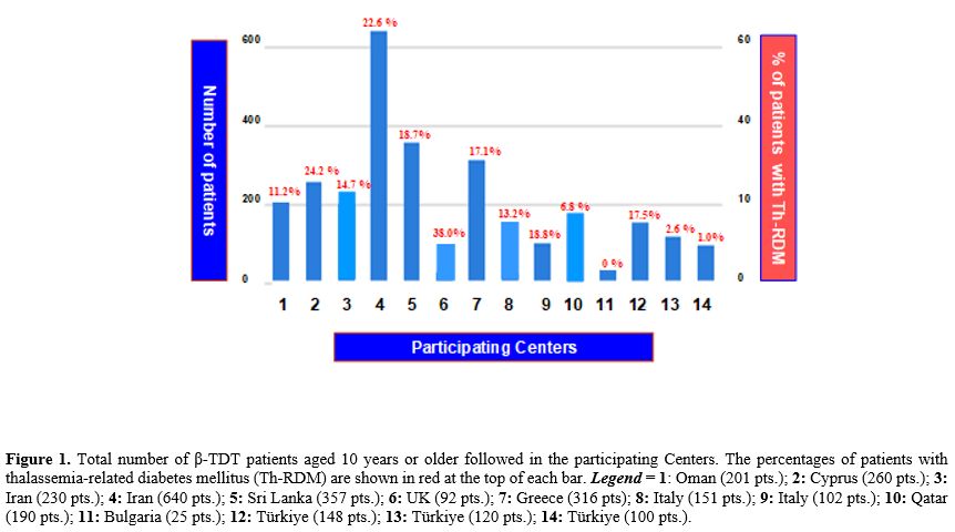
Survey response rate. 14 out of 18 invited Centers (Bulgaria [1], Cyprus [1], Greece [1], Iran [2], Italy [2], Oman [1], Qatar [1], Sri Lanka [1], Türkiye [3], United Kingdom [1]) accepted the invitation to take part in the survey, a response rate of 77.7%.Centers’ characteristics. The total number of β-TDT patients followed at the participating Centers was 3,372, of whom 2,932 (86.9%) were aged 10 years or older (Figure 1). Twelve of 14 Centers (85.7%) monitor more than 100 β-TDT patients; nine (64.2%) follow both pediatric and adult β-TDT patients, and 10 (71.4%) are affiliated with an academic institution.
 |
|
The total number of Th-RDM reported by all Centers was 549; all patients were aged 10 years or older. The overall mean prevalence of Th-RDM was 14.7 ± 10.2% (range: 0% - 38.0%; median: 15.9%). No patient with Th-RDMs was reported by a pediatric Center, whereas 25 β-TDT patients over the age of 10 were reported (Figure 1, no. 11).
Notably, the survey did not include patients' age, the year of Th-RDM diagnosis, or the use of HbA1c for the diagnosis of dysglycemia. The latter was not included due to poor sensitivity in diagnosing dysglycemia in β-TDT patients, as defined by the current diagnostic threshold.[12]
Screening and criteria used for the diagnosis of dysglycemia. All Centers routinely checked one of the following every 1-12 months (median 7.5 months): fasting PG (10/14; 71.4%); fasting serum glucose (3/14; 21.4%); and capillary blood glucose (1/14; 7.1%). OGTT is recommended by 10 Centers, with a mean age at initiation of 13 ± 2.8 years (range: 10-18 years). In the remaining 3 Centers, OGTT is requested based on fasting serum glucose results.
The OGTT was carried out at the thalassemia Centers in 5/14 (35.7%), at the hospital biochemistry laboratories in 6/14 (42.8%), and at both thalassemia Centers and private biochemistry laboratories in 3/14 (21.4%).
The most commonly reported time points for OGTT screening were fasting and 2 h post-glucose challenge, or fasting, 60, and 120 minutes. post glucose challenge test (8/14 Centers; 57.1 %). In the remaining 4 Centers, the number of blood glucose samples collected during OGTT was 4 (2/14; 14.2%), 5 (1/14; 5.8%), and 6 (1/14; 5.8%). In 2 centers, the screening was mainly based on FPG. In 5 out of 14 Centers (35.7%), the OGTT was performed on the same day as blood transfusion(s), and in the remaining 9 Centers, within a few days after blood transfusion(s). The amount of glucose solution (anhydrous, freshly prepared or ready-to-use solution) given for OGTT was 210 ± 55 ml (median 225 ml, range:110-300 ml).
Hexokinase or glucokinase enzyme analysis was used in ~93% of laboratories for glucose measurement. In one Center, capillary blood from the fingertip was used to measure glucose. Five Centers (35.7%) also determined insulin levels during the OGTT. These additional measurements enabled assessment of insulin sensitivity and secretion, as well as β-cell function. Dysglycemia is classified according to the ADA criteria 4 by 9 Centers (64.2%) and in the remaining Centers by WHO criteria.[5] Both organizations define diabetes mellitus (DM) as: (a) a fasting PG of ≥126 mg/dL (≥ 7.0 mmol/L), or (b) a 2-hour PG ≥ 200 mg/dL (≥ 11.1 mmol/L), during oral glucose tolerance test (OGTT), or (c) a random PG ≥ 200 mg/dL (≥ 11.1 mmol/L) with classic diabetes symptoms. In the absence of unequivocal hyperglycemia, the ADA[4] recommends confirming the result with repeat testing. In symptomatic hyperglycemia, the diagnosis is obvious, and a confirmatory test is not required before treatment is initiated. However, when results from more than one test are available and discordant, the test(s) should be repeated. In patients with impaired fasting glucose (IFG) or impaired glucose tolerance (IGT), glucose homeostasis is reassessed on average after 6-8 months (range: 1-12 months) (median 6 months in 11/13 Centers). In the remaining 3 Centers, the reassessment is based on fasting serum glucose levels.
According to the criteria of the International Diabetes Federation Position Statement[13] for diagnosing hyperglycemia, 1-hour post-load PG≥ 155 mg/dL (≥8.6 mmol/L) is considered an early biomarker of dysglycemia and is associated with poorer β-cell function and lower insulin sensitivity.[13] This additional criterion is utilized by 8 out of 14 Centers (57.1%). Two Centers reported that patients must pay out of pocket for the OGTT. For this reason, at one Center, OGTT was required only if fasting serum glucose levels were abnormal.
Preanalytical phase and quality of indicators: Patients' preparation and sampling. The quality of information and instructions to patients with regard to preparation for OGTT was arbitrarily classified as: (i) full agreement with ADA[4] and WHO[5] recommendations (6/14; 42.8 %), (ii) partial agreement with recommendations (6/14; 42.8 %), and (iii) lack of structured information (2/14;14.2%). Fully recommended information included: a preparatory diet, water intake, advice on fasting duration (> 8 hours), and restrictions prior to OGTT (caffeine, smoking, and vigorous physical activity). Moreover, factors that may influence test results (e.g., medication such as thiazide diuretics, beta-blockers, and corticosteroids; recent infection, etc.) were recorded and, in case of non-adherence, the OGTT was postponed. In all thalassemia Centers, OGTT was carried out after a recommended overnight fasting of 10 ± 2 hours (range:8-14 hours), using 1.75 g/kg (max.75 g of glucose solution).
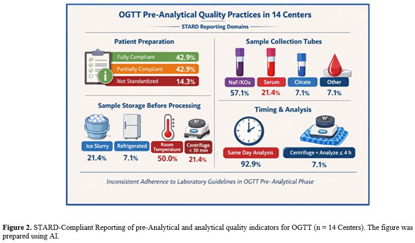
The application of STARD-aligned quality indicators indicates incomplete standardization of the OGTT pre-analytical phase across participating centers. Although most laboratories performed same-day analysis, key procedural elements — such as immediate centrifugation, cold-chain preservation, and standardized patient preparation — were inconsistently implemented. Notably, half of the centers stored samples at room temperature prior to processing, a practice that may introduce glycolysis-related bias and affect diagnostic accuracy. Limited reporting of exact transport and processing intervals further reduces reproducibility. These findings indicate variability in adherence to recommended laboratory standards and highlight the need for harmonized pre-analytical protocols (Figure 2).
 |
|
In summary, the graphical abstract clearly synthesizes the multicenter ICET-A findings, emphasizing how variability in patient preparation and sample handling during the pre-analytical phase can significantly affect OGTT accuracy in β-TDT. By visually linking laboratory instability (glycolysis, tube selection, transport delays) to potential diagnostic misclassification, it effectively highlights the urgent need for harmonized STARD-aligned protocols to improve the reliability of dysglycemia screening across Centers.
The interval time from sample collection to centrifugation and analysis. The precise interval time from venepuncture to the arrival of the blood sample to laboratories was not available; in general, PG or serum glucose levels were analyzed on the same day of testing, with one exception, where the specimens were analyzed for serum glucose determination within 4 hours.
Discussion
The total testing process (TTP) for OGTT comprises three phases: pre-analytical, analytical, and post-analytical. The impact of pre-analytical factors on the blood glucose measurements is very important not only for the diagnosis of diabetes but also for identifying high risk patients and to assessing the efficacy of different iron chelation regimes.The International Organization for Standardization (ISO 15189:2022) of accredited laboratories has defined the pre-analytical phase as "the steps that consider the healthcare personnel’s request, patient preparation, sample collection, transportation to and within the laboratory, ending with the start of the analysis process. It is the most important phase of TTP that requires standardized procedures to minimize variability and bias, both in terms of required analytical methods and biological variability".[14] The importance of the pre-analytical phase is often underestimated and accounts for 46–68% of all laboratory errors.[15]
Therefore, given the narrow diagnostic thresholds used to define dysglycemia, even modest pre-analytical deviations may lead to clinically meaningful misclassification, with potential adverse outcomes and increased healthcare costs.
By documenting current practices across ICET-A Centers, this study provides a pragmatic, descriptive framework to improve standardization and diagnostic reliability in β-TDT care. Substantially, our survey has shown that adherence to, and deviations from, current international guidelines[4,5] are variable and have important implications for clinical practice. The lowest adherence rate concerned the information and instructions given to patients prior to OGTT and the storage of serum or plasma glucose samples from collection to centrifugation and analysis. Unfortunately, it is difficult to guarantee the immediate delivery of blood samples to the laboratory nowadays. It has become routine to send uncentrifuged samples several hours after collection, which further impairs glucose stability and contributes to errors in the diagnosis of dysglycemia. However, compliance with this recommendation is particularly challenging for the OGTT, as fasting and post-glucose samples are usually held at the point of patient care until the test is completed over 2 h.[6,16]
There are two main approaches to inhibit glycolysis. The first, known for many years, consists of placing the blood tube in an ice–water slurry immediately after blood collection and separating the plasma from the cells within 30 minutes.[7] However, it would be highly impractical to expect that every sample would be cooled and separated soon after collection, and placing tubes at 4°C marginally reduces the decay of PG concentration.[17] The second approach involves collecting venous whole blood into tubes containing a glycolytic inhibitor.[6,16] Traditionally, sodium fluoride (NaF) used alone or, more commonly, with anticoagulants like potassium oxalate, has been used as a long-term glucose stabilizer but it does not exert immediate inhibition and may require up to 3-4 h to fully suppress enolase glycolytic enzyme activity.[18] Gambino et al.[19] have reported a reduction of PG concentration of 4.6% at 2-h and by 7.0% at 24 h when blood was drawn into tubes containing a combination of sodium fluoride (NaF) and potassium oxalate (KOx). To address this limitation, citrate-buffered tubes have been developed to enhance glycolysis inhibition by lowering sample pH. Acidification inhibits hexokinase and phosphofructokinase, enzymes that act early in the glycolytic pathway. The inhibitory effect of acidification is sustainable for approximately 10 h at 25°C. However, it should be noted that although citrate tubes maintain long-term glucose stability at room temperature, the rates of decline of glucose in the first hour after sample collection in tubes with and without fluoride are virtually identical.[18] Furthermore, some observational studies have shown that the shift from NaF to citrate-buffered tubes has contributed to an over-estimation (~13% ) in PG measurements when compared to blood collected into conventional tubes under cooled conditions.[20] An additional problem of citrate-buffered tubes is the lack of standardization and clinically significant biases between different tube types used for glucose measurement.[21-23] Therefore, it has been suggested that, before a wider adoption of citrate-buffered tubes, larger studies are needed to confirm or redefine current diabetes diagnostic thresholds.[24-26] Waiting for the latest findings in this field, the benefits of fluoride-citrate induced glucose stabilization are likely to outweigh the disadvantage of a small over-estimation in measured PG, when compared to other collection tube systems.[27]
Future priorities
The significant heterogeneity in preanalytical factors affecting glucose measurement across participating Thalassemia Centers raises the question of whether this could result in a significant difference in the reported prevalence of dysglycemia. This survey cannot address this issue, but it highlights the need for studies aimed at examining the glycemic status of patients in these Centers by applying the best available practices in preparation, collection, handling, and analysis, and comparing the results with the reported rates.Of note, the utility of HbA1c as a criterion for diagnosis of dysglycemia is limited in people with thalassemia major due to the impact of anemia requiring regular red cell transfusions. For this reason, other biomarkers, such as fructosamine and glycated albumin, have been used for monitoring of glycemic control in these patients, without having been validated as screening tests for glycemia in this population.[28-33] Therefore, OGTT remains the gold standard test for screening of glycemia in patients with thalassemia, although its value has been questioned due to pre-analytical, analytical and post-analytical variables affecting its reproducibility and accuracy.[6,8] Patients with plasma glucose levels closer to cut-off values require more attention to avoid diagnostic misclassification.
Limitations
OOur study survey has several limitations, the first being the limited number of Centers participating in the ICET-A survey. Nevertheless. The total number of β-TDT patients followed in the participating Centers was large. Second, most of the detected problems were based on self-reported responses from different Centers. Further, the survey did not include an evaluation of patients’ adherence to recommended information, which could be important for the impact of guidelines. Thirdly, the variability of answers reported by the Centers on specific pre-analytic questions reported in the questionnaire and the limited number of subgroups of patients did not permit a comparison of performance between hematologists versus endocrinologists, adherence level between academic vs. non-academic Centers, pediatric vs. mixed adult–pediatric centers, and magnitude of misclassification attributable to pre-analytical deviations. Finally, the distance, time, and mode of transport between the Centers/Clinics where blood was drawn and the central laboratory, and the time required for blood glucose determinations were not included in the questionnaire.Other aspects remain poorly explored; for example, very little is known about the acute effects of blood transfusion on glucose homeostasis in β-TDT patients. Wankanit et al.[34] have reported that an increase in Hb of about 1.5 g/dL, after acute blood transfusions, caused an increase of insulin secretion and a reduced insulin sensitivity, secondary to an increase in serum ferritin level (~ 400 µg/L).
Conclusions
Based on the Standards for Reporting of Diagnostic Accuracy (STARD) statement, the pre-analytical blood sampling procedures for OGTT screening in thalassemia Centers involved in the survey require revision and standardization, recognizing that they can be controlled and limited but cannot be fully eliminated.Finally, we would like to suggest: (i) collaboration between hematologists, endocrinologists, diabetologists, biochemists, nurses and technicians to increase the awareness oflaboratory variability in each single step, described above with the aim of improving the reliability of the results, and to advice (ii) researchers to report, in their scientific publications, the methods used in the preanalytical phase for a better interpretation and comparison of data on glucose homeostasis between different Centers.
Author Contributions
VDS and SD designed and implemented the survey. VDS, SD, and PT wrote the manuscript. ATS, DC, and CK contributed to discussions and reviewed/edited the manuscript. ATS performed the STARD analysis and prepared Figure 2. All Centers certify that they have participated in the work to take public responsibility for the content, including presentation of data reported in the survey. VDS takes responsibility for the accuracy of data analysis. All authors read and approved the final version of the manuscript.References
- De Sanctis V, Soliman AT, Elsedfy H, AL Yaarubi S,
Skordis N, Khater D, El Kholy M, Stoeva I, Fiscina B, Angastiniotis M,
Daar S, Kattamis C. The ICET-A recommendations for the diagnosis and
management of disturbances of glucose homeostasis in thalassemia major
patients. Mediterr J Hematol Infect Dis. 2016; 8 (1):e2016058. https://doi.org/10.4084/mjhid.2016.058 PMid:27872738 PMCid:PMC5111521
- De
Sanctis V, Canatan D, Daar S, Kattamis C. (Steering Committee); Banchev
A, Modeva I, Savvidou I, Christou S, Kattamis A, Delaporta P,
Kostaridou-Nikolopoulou S, Karim M, Saki F, Faranoush M, Campisi S,
Fortugno C, Gigliotti F, Wali Y, Al Yaarubi S, Yassin MA, Soliman AT,
Kottahachchi D, Kurtoğlu E, Gorar S, Turkkahraman D, Unal S, Oymak Y,
Ay Tuncel D, Karakas Z, Gül N, Yildiz M, Elhakim I, Tzoulis P.
(Participants). A multicenter ICET-A survey on adherence to annual oral
glucose tolerance test (OGTT) screening in transfusion-dependent
thalassemia (TDT) patients -The expert clinicians' opinion on factors
influencing the adherence and on alternative strategies for adherence
optimization. Mediterr J Hematol Infect Dis. 2025, 17(1): e2025008. https://doi.org/10.4084/MJHID.2025.008 PMid:39830799 PMCid:PMC11740908
- Tzoulis
P, Yavropoulou MP, Banchev A, Modeva I, Daar S, De Sanctis V. Recent
advancements in glucose dysregulation and pharmacological management of
osteoporosis in transfusion-dependent thalassemia (TDT): an update of
ICET-A (International Network of Clinicians for Endocrinopathies in
Thalassemia and Adolescence Medicine). Acta Biomed. 2023;94(3)3:e2023178. https://doi.org/10.23750/abm.v94i3.14805
- American
Diabetes Association Professional Practice Committee; 2. Diagnosis and
Classification of Diabetes: Standards of Care in Diabetes-2025.
Diabetes Care. 2025; 48 (Supplement 1): S27-S49. https://doi.org/10.2337/dc25-S002 PMid:39651986 PMCid:PMC11635041
- World Health Organization. Classification of diabetes mellitus. Geneva: World Health Organization. 2019. p.1-36. https://iris.who.int/server/api/core/bitstreams/2cb3ab68-a52a-402e-ad47-8bc5a4edc834/content
- Bogdanet
D, O'Shea P, Lyons C, Shafat A, Dunne F. The Oral Glucose Tolerance
Test-Is It Time for a Change?-A Literature Review with an Emphasis on
Pregnancy. J Clin Med. 2020; 9 (11):3451. https://doi.org/10.3390/jcm9113451 PMid:33121014 PMCid:PMC7693369
- Sacks
DB, Arnold M, Bakris GL, Bruns DE, Horvath AR, Lernmark Å, Metzger BE,
Nathan DM, Kirkman MS. Guidelines and Recommendations for Laboratory
Analysis in the Diagnosis and Management of Diabetes Mellitus. Diabetes
Care. 2023;46(10):e151-e199. https://doi.org/10.2337/dci23-0036 PMid:37471273 PMCid:PMC10516260
- Mshelia
DS, Adamu S, Galli RM. Oral glucose tolerance test (OGTT): Undeniably
the first choice investigation of dysglycaemia, reproducibility can be
improved.In: Type 2 Diabetes - From Pathophysiology to Cyber Systems.
Intech Open. Ed.Anca Pantea Stoian. 2021:1-38. http://doi.org/10.5772/intechopen.96549
- Nikolac
N, Lenicek Krleza J, Simundic AM. Preanalytical external quality
assessment of the Croatian Society of Medical Biochemistry and
Laboratory Medicine and CROQALM: finding undetected weak spots.
Biochemia Medica. 2017;27(1):131-143. https://doi.org/10.11613/BM.2017.017 PMid:28392736 PMCid:PMC5382847
- Isabel
García-del-Pino , Josep M. Bauça1, Carolina Gómez, Andrea Caballero ,
María Antonia Llopis, Mercedes Ibarz , Débora Martínez, Montserrat
Ventura, Itziar Marzana, Juan J. Puente1, Marta Segovia, Paloma Salas,
Rubén Gómez-Rioja. Preanalytical issues related to routine and
diagnostic glucose tests: Results from a survey in Spain. Biochem Med
(Zagreb) 2020;30(1):010704. https://doi.org/10.11613/BM.2020.010704 PMid:31839724 PMCid:PMC6904967
- Cohen
JF, Korevaar DA, Altman DG, Bruns DE, Gatsonis CA, Hooft L, Irwig L,
Levine D, Reitsma JB, de Vet HC, Bossuyt PM. STARD 2015 guidelines for
reporting diagnostic accuracy studies: explanation and elaboration. BMJ
Open. 2016;6(11):e012799. https://doi.org/10.1136/bmjopen-2016-012799 PMid:28137831 PMCid:PMC5128957
- Mahmoud
AA, El-Hawy MA, Allam ET, Salem AH, Hola AS. HbA1c or fructosamine on
evaluating glucose intolerance in children with beta- thalassemia.
Pediatr Res.2024. https://doi.org/10.1038/s41390-024-03146-y PMid:38575692 PMCid:PMC11521987
- Bergman
M, Manco M, Satman I, Chan J, Schmidt MI, Sesti G, Vanessa Fiorentino
T, Abdul-Ghani M, Jagannathan R, Kumar Thyparambil Aravindakshan P,
Gabriel R, Mohan V, Buysschaert M, Bennakhi A, Pascal Kengne A, Dorcely
B, Nilsson PM, Tuomi T, Battelino T, Hussain A, Ceriello A, Tuomilehto
J. International Diabetes Federation Position Statement on the 1-hour
post-load plasma glucose for the diagnosis of intermediate
hyperglycaemia and type 2 diabetes. Diabetes Res Clin Pract.
2024;209:111589. https://doi.org/10.1016/j.diabres.2024.111589 PMid:38458916
- International
Organization for Standardization. ISO 15189 - Medical Laboratories
Requirements for Quality and Competence. 4th ed. Geneva: ISO, Ed., 2022
- Plebani M. The detection and prevention of errors in laboratory medicine. Ann ClinBiochem. 2010; 47:101-110. https://doi.org/10.1258/acb.2009.009222 PMid:19952034
- Zubair M, Launico MV. Glucose tolerance test [Updated 2025 Sept.15]. In: Statpearls [Internet] Available from: https://www.ncbi.nlm.nih.gov/sites/books/NBK532915/
- Nevraumont
A, Deltombe M, Bayart J L. How pre-analytical conditions impact glucose
measurement and (gestational) diabetes diagnosis: A real-world
stability study and a call for harmonization. Clin Chim Acta. 2024;562:
119875. https://doi.org/10.1016/j.cca.2024.119875 PMid:39013524
- Balboni F, Burbui S, Lippi G. Glucose variation in centrifuged serum and lithium-heparin gel tubes stored for up to 96 hours at room temperature or 4 °C. Scand J Clin Lab Invest. 2018;78:546-550. https://doi.org/10.1080/00365513.2018.1517221 PMid:30755098
- Gambino
R, Piscitelli J, Ackattupathil TA,Theriault JL, Andrin RD, Sanfilippo
ML, Etienne M. Acidification of blood is superior to sodium fluoride
alone as an inhibitor of glycolysis, Clin Chem. 2009; 55 (5):1019-1021,
https://doi.org/10.1373/clinchem.2008.121707 PMid:19282354
- Nevraumont
A, Deltombe M, Bayart JL. How pre-analytical conditions impact glucose
measurement and (gestational) diabetes diagnosis: A real-world
stability study and a call for harmonization. Clin Chim Acta.
2024;562:119875. https://doi.org/10.1016/j.cca.2024.119875 PMid:39013524
- Gupta
S, Kaur H.Inhibition of Glycolysis for Glucose Estimation in Plasma:
Recent Guidelines and their Implications. Ind J Clin Biochem.
2014;29(2):262-264. https://doi.org/10.1007/s12291-013-0405-1 PMid:24757316 PMCid:PMC3990791
- Grzych
G, Defauwes I, de Tullio P, Pekar JD, Brousseau T, Lippi G, Cavalier E.
Blood glucose measurement inside and outside the laboratory: both
preanalytical and analytical challenges. Crit Rev Clin Lab Sci.
2025;62(8):613-630. https://doi.org/10.1080/10408363.2025.2533855 PMid:40859885
- Pasqualetti
S, Braga F, Panteghini M. Pre-analytical and analytical aspects
affecting clinical reliability of plasma glucose results. Clin Biochem.
2017;50(10-11):587-594. https://doi.org/10.1016/j.clinbiochem.2017.03.009 PMid:28300544
- Saracevic
A, Dukic L, Juricic G, Milevoj Kopcinovic L, Mirosevic G, Simundic AM.
Various glycolysis inhibitor-containing tubes for glucose measurement
can not be used interchangeably due to clinically unacceptable biases
between them. Clin Chem Lab Med. 2018;56:236-241. https://doi.org/10.1515/cclm-2017-0279 PMid:28746044
- Bonetti
G, Giavarina D, Carta M. Clinical impact of citrate- containing tubes
on the detection of glucose abnormalities by the oral glucose tolerance
test. Diagnosis (Berl). 2019;6:377-383. https://doi.org/10.1515/dx-2018-0100 PMid:30875321
- Pasqualetti
S, Panteghini M. Clinical impact of glycolysis inhibition on plasma
glucose results requires caution. Ann Clin Biochem. 2017;54:302-303. https://doi.org/10.1177/0004563216659091 PMid:27343222
- Rebekah
Carey R, Lunt H, Heenan HF, Frampton CMA, Florkowski CM. Collection
tubes containing citrate stabiliser over-estimate plasma glucose, when
compared to other samples undergoing immediate plasma separation. Clin
Biochem.2016; 49:1406-1411. https://doi.org/10.1016/j.clinbiochem.2016.05.017 PMid:27234599
- De
Sanctis V, Soliman AT, Elsedfy H, Yaarubi SA, Skordis N, Khater D, El
Kholy M, Stoeva I, Fiscina B, Angastiniotis M, Daar S, Kattamis C. The
ICET-A Recommendations for the Diagnosis and Management of Disturbances
of Glucose Homeostasis in Thalassemia Major Patients. Mediterr J
Hematol Infect Dis. 2016;28;8(1):e2016058. https://doi.org/10.4084/mjhid.2016.058 PMid:27872738 PMCid:PMC5111521
- Gomber
S, Bagaria A, Madhu SV, Dewan P. Glucose Homeostasis Markers in
Beta-Thalassemia. J Pediatr Hematol Oncol. 2018;40(7):508-510. https://doi.org/10.1097/MPH.0000000000001161 PMid:29668553
- Candrarukmi
D, Annang GM,Muhammad R. Glycated albumin as marker for early
hyperglycemia detection in adolescent with β thalassemia major.Indones
Biomed J. 2021;13(3): 281-288. ISSN: 2085-3297.Online ISSN: 2355-9179 https://doi.org/10.18585/inabj.v13i3.1546
- Paroni
R, Ceriotti F, Galanello R, Battista Leoni G, Panico A, Scurati E,
Paleari R, Chemello L, Quaino V, Scaldaferri L, Lapolla A, Mosca A.
Performance characteristics and clinical utility of an enzymatic method
for the measurement of glycated albumin in plasma. Clin Biochem.
2007;40(18):1398-405. https://doi.org/10.1016/j.clinbiochem.2007.08.001 PMid:17919531
- Mahmoud
AA, El-Hawy MA, Allam ET, Salem AH, Abo Hola AS. HbA1c or fructosamine
on evaluating glucose intolerance in children with beta- thalassemia.
Pediatr Res. 2024;96(5):1292-1298. https://doi.org/10.1038/s41390-024-03146-y PMid:38575692 PMCid:PMC11521987
- Delaporta
P, Bistas K, Chatzikali E, Maravgaki V, Agiomavrititephanopoulou E,
Chouliara V, Kyriakopoulou D, Berdalli S, Kattamis A. Oral glucose
tolerance test remains the gold standard for diagnosis of glucose
metabolism disturbances in thalassemia patients. Blood.2025;146
(Supplement 1):1144. https://doi.org/10.1182/blood-2025-1144
- Wankanit S, Chuansumrit A, Poomthavorn P, Khlairit P, Pongratanakul S, Mahachoklertwattana P. Acute Effects of Blood Transfusion on Insulin Sensitivity and Pancreatic β-Cell Function in Children with β-Thalassemia/Hemoglobin E Disease. J Clin Res Pediatr Endocrinol. 2018;10(1):1-7. https://doi.org/10.4274/jcrpe.4774 PMid:28739553 PMCid:PMC5838366