 |
|
Case 1: young patient with progressive MCL
Case 2: life-threatening GvHD
A 59-year-old male patient was diagnosed with ASM in January 2018. He had a previous diagnosis of tubular adenoma of the colon with low-grade dysplasia and Helicobacter pylori (Hp) related chronic gastritis. Since 2014, he has reported grade II neutropenia for which he has performed periodic blood count analysis. In December 2017, he presented to the ER with persistent fever: physical examination revealed splenomegaly confirmed by US evaluation (spleen 18 cm), and laboratory tests showed grade II anemia and grade IV neutropenia. Bone marrow evaluation revealed 25% infiltration by a a multifocal cluster of atypical CD2-/CD25+ MCs and grade I fibrosis, together with the presence of the KIT D816V mutation; serum tryptase was 100 mcg/L. In September 2018, he started midostaurin 100 mg twice daily, with limited benefit; in November 2020, bone marrow evaluation showed 40% MC infiltration and grade II fibrosis; concomitant serum tryptase was 132 mcg/L. Endoscopy revealed colon and gastro-duodenal infiltration of MCs (Figure 1, A to D), together with Hp infection. Cladribine 0.14 mg/kg daily infusion for 5 consecutive days was initiated; HLA typing and family donor screening were also performed. In March 2021, he presented to the ER with fever, nausea, vomiting, and scrotal abscess: he received multiple courses of antibiotic therapy and red blood cell transfusions. In July 2021, he underwent an allogeneic transplant from a haploidentical donor: bone marrow evaluation showed 75% MCs infiltration and grade III fibrosis. The pre-engraftment period was complicated by suspected veno-occlusive disease, diarrhea, and fever, which resolved after defibrotide and empiric antibiotic therapy. In August 2021, bone marrow evaluation detected a 25% MCs infiltration with persistent KIT D816V and grade II fibrosis. In September 2021, he came to the ER because of worsening diarrhea and vomiting, suspicious for intestinal GvHD. After a negative microbiological workup, extracorporeal photopheresis, steroids, and ruxolitinib were employed. Bone marrow evaluation revealed persistent 30% MCs infiltration and grade II fibrosis, and gastro-intestinal biopsies detected lamina propria inflammation suggestive of GvHD but no MCs infiltration or Hp infection (Figure 1, E-F). He experienced progressive worsening of clinical conditions after the onset of pleural effusion, hypotension requiring amine support, and abdominal pain with bloody vomiting. He was transferred to the intensive care unit (ICU) and died a few days later in November 2021 from progressive deterioration.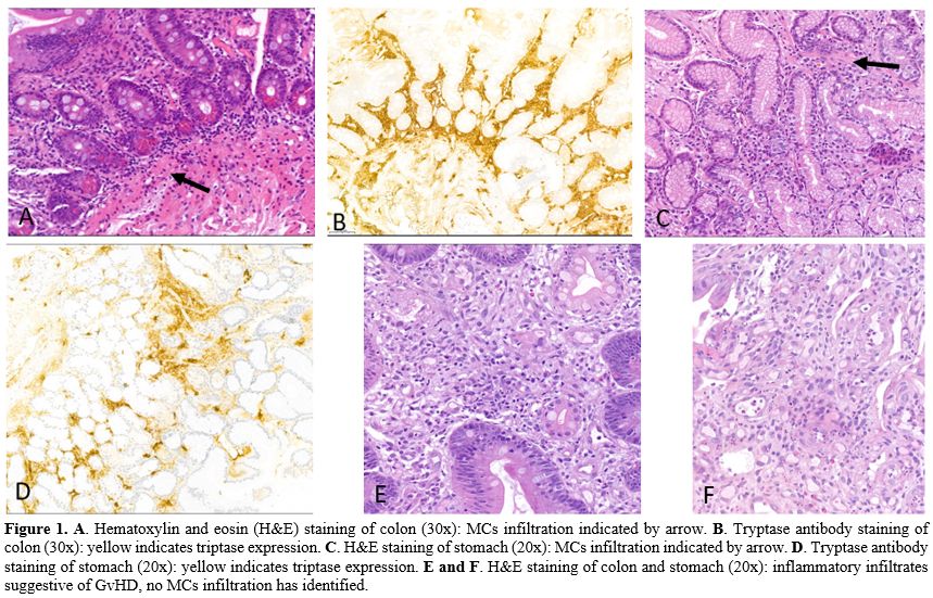 |
Figure 1. A. Hematoxylin and eosin (H&E) staining of colon (30x): MCs infiltration indicated by arrow. B. Tryptase antibody staining of colon (30x): yellow indicates triptase expression. C. H&E staining of stomach (20x): MCs infiltration indicated by arrow. D. Tryptase antibody staining of stomach (20x): yellow indicates triptase expression. E and F. H&E staining of colon and stomach (20x): inflammatory infiltrates suggestive of GvHD, no MCs infiltration has identified. |
Case 3: late recovery of bone marrow function
Conclusions and Future Perspectives
Comparing the three most considerable studies about transplant in AdvSM, OS, and PFS after transplant were largely overlapping: at 3 years, about 40-50% of patients progressed, and almost 50% died.[6,10,11] Some considerations could be discussed. First, the study's population includes mostly patients with SM-AHN, among whom the associated hematological neoplasm is often responsible for the indication to transplant. Different authors have reported a reduced efficacy of graft-versus-mast cell effect compared to other hematopoietic compartments. This is particularly prominent in extra-medullary disease, irrespective of the intensity of conditioning regimens,[6,10] as reported for most myeloid neoplasms.[12] Furthermore, single case reports have shown a delayed clearance of bone marrow MCs infiltration.[13,14] Second, the time of observation is wide and not directly comparable to today, when TKI are widely available for AdvSM treatment. Nevertheless, recent changes in transplant management have improved non-relapsed mortality and mitigated the severity of GvHD.
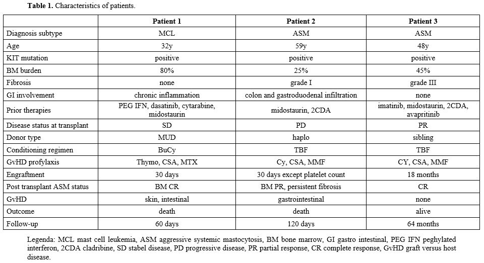
According to clinical conditions and age, the transplant option should be discussed early with patients to plan HLA typing and familiar/unrelated donor screening. Myeloablative conditioning (MAC) regimens are known to be highly effective in disease control, although a higher rate of drug toxicity and increased GvHD severity have been observed. In the last few years, the age limit for the procedure has progressively increased, following the introduction of reduced-intensity conditioning (RIC) regimens. A worse survival was reported following RIC among 57 patients transplanted between 1990 and 2013,[6] although RIC was used in patients with lower performance status. More recently, a retrospective analysis of 71 patients transplanted between 1999 and 2021 could not conclusively establish the superiority of one conditioning regimen over another, likely due to the wide range of regimens used.[10] In our patients, the use of MAC regimens obtained disease control in all cases and timely engraftment in 2/3 cases, while severe GvHD was fatal for two patients.
Transplant should be performed when the best disease control has been achieved: lack of response was reported to predict an adverse prognosis among 71 patients transplanted between 1999 and 2021.[10] In our third patient, the use of avapritinib before transplant reduced the burden of disease, while grade III fibrosis persisted at the time of transplant: the presence of fibrosis since diagnosis may have impaired bone marrow recovery irrespective of conditioning regimens, as for other myeloid neoplasms.[15]
In our case series, we reported a good tolerability of MAC with valuable disease eradication and variable timing of engraftment. A potential difficulty in GvHD control has emerged, especially if the target organ has been a site of disease involvement: prior GI mast-cell involvement may warrant careful pre-transplant assessment, as severe post-transplant GI GvHD/toxicity may be difficult to control. The limitations of this report are related to the small number of cases and the relatively long observation period, during which both therapeutic options and transplant procedures have advanced. More data are warranted to better understand the potential correlation between MC infiltration and the severity of GvHD.
In this case series, two patients reported symptoms predictive of MCs' infiltration of the GI tract at the onset of AdvSM. In the post-engraftment phase, both developed acute severe GI GvHD, irrespective of conditioning regimen and GvHD prophylaxis. Acute GvHD has been reported in 42% of patients in two studies,[6,10] while it was about 20% after wider use of RIC in a more recent study.[11] No apparent correlation with prior disease infiltration has been reported so far. Recently, different studies have identified elevated MCs infiltration in the GI tract in patients with unexplained chronic diarrhea, disorders of gut-brain interaction, and irritable bowel syndrome.[16,17,18] MC activation causes hypersecretion, alterations in intestinal motility, visceral hypersensitivity, and alterations in the gut epithelial barrier through the release of inflammatory mediators such as histamine, prostaglandins, serotonin, and tryptase.[19,20] Taken together, these observations may suggest a contributory role for mediators released from MCs after the graft-versus-mast cell effect, increasing local toxicity and ultimately causing damage. In patients with AdvSM, pre-transplant GI involvement may be a clinical warning sign requiring careful assessment and monitoring.
Acknowledgments
There are no acknowledgments to report.Footnotes
The study was conducted in accordance with the Declaration of Helsinki. Ethical review and approval were waived for this study due to report of a case series with no formal analysis aiming to evaluate the efficacy of a therapeutic approach.Informed consent to publication was obtained verbally from living patients and recorded in the medical record.
Author contributions
CM Conceptualization; Data curation; Writing – original draft. FL, CP, and SS Data curation; Writing – review & editing. PL Conceptualization; Data curation; Writing – review & editing.Availability of data and materials
All data reported in the manuscript are available on request.References
- Valent P, Akin C, Metcalfe DD. Mastocytosis: 2016
updated WHO classification and novel emerging treatment concepts. Blood
2017:129, 1420-1427. https://doi.org/10.1182/blood-2016-09-731893 PMid:28031180 PMCid:PMC5356454
- Arber
DA, Orazi A, Hasserjian R, Thiele J, Borowitz MJ, Le Beau MM,
Bloomfield CD, Cazzola M, Vardiman JW. The 2016 revision to the World
Health Organization classification of myeloid neoplasms and acute
leukemia. Blood 2016; 127:2391-2405. https://doi.org/10.1182/blood-2016-03-643544 PMid:27069254
- Khoury
JD, Solary E, Abla O, Akkari Y, Alaggio R, Apperley JF, Bejar R, Berti
E, Busque L, Chan JKC, Chen W, Chen X, Chng WJ, Choi JK, Colmenero I,
Coupland SE, Cross NCP, De Jong D, Elghetany MT, Takahashi E, Emile JF,
Ferry J, Fogelstrand L, Fontenay M, Germing U, Gujral S, Haferlach T,
Harrison C, Hodge JC, Hu S, Jansen JH, Kanagal-Shamanna R, Kantarjian
HM, Kratz CP, Li XQ, Lim MS, Loeb K, Loghavi S, Marcogliese A,
Meshinchi S, Michaels P, Naresh KN, Natkunam Y, Nejati R, Ott G, Padron
E, Patel KP, Patkar N, Picarsic J, Platzbecker U, Roberts I, Schuh A,
Sewell W, Siebert R, Tembhare P, Tyner J, Verstovsek S, Wang W, Wood B,
Xiao W, Yeung C, Hochhaus A. The 5th edition of the World Health
Organization Classification of Haematolymphoid Tumours: Myeloid and
Histiocytic/Dendritic Neoplasms. Leukemia. 2022;36(7):1703-1719. https://doi.org/10.1038/s41375-022-01613-1 PMid:35732831 PMCid:PMC9252913
- Arber
DA, Orazi A, Hasserjian RP, Borowitz MJ, Calvo KR, Kvasnicka HM, Wang
SA, Bagg A, Barbui T, Branford S, Bueso-Ramos CE, Cortes JE, Dal Cin P,
DiNardo CD, Dombret H, Duncavage EJ, Ebert BL, Estey EH, Facchetti F,
Foucar K, Gangat N, Gianelli U, Godley LA, Gökbuget N, Gotlib J,
Hellström-Lindberg E, Hobbs GS, Hoffman R, Jabbour EJ, Kiladjian JJ,
Larson RA, Le Beau MM, Loh ML, Löwenberg B, Macintyre E, Malcovati L,
Mullighan CG, Niemeyer C, Odenike OM, Ogawa S, Orfao A, Papaemmanuil E,
Passamonti F, Porkka K, Pui CH, Radich JP, Reiter A, Rozman M, Rudelius
M, Savona MR, Schiffer CA, Schmitt-Graeff A, Shimamura A, Sierra J,
Stock WA, Stone RM, Tallman MS, Thiele J, Tien HF, Tzankov A, Vannucchi
AM, Vyas P, Wei AH, Weinberg OK, Wierzbowska A, Cazzola M, Döhner H,
Tefferi A. International Consensus Classification of Myeloid Neoplasms
and Acute Leukemias: integrating morphologic, clinical, and genomic
data. Blood. 2022;140(11):1200-1228. https://doi.org/10.1182/blood.2022015850 PMid:35767897 PMCid:PMC9479031
- Pardanani
A, Reichard K, Tefferi A. Advanced systemic mastocytosis-Revised
classification, new drugs and how we treat. Br J Haematol.
2024;204(2):402-414. https://doi.org/10.1111/bjh.19245 PMid:38054381
- Ustun
C, Reiter A, Scott BL, Nakamura R, Damaj G, Kreil S, Shanley R, Hogan
WJ, Perales MA, Shore T, Baurmann H, Stuart R, Gruhn B, Doubek M, Hsu
JW, Tholouli E, Gromke T, Godley LA, Pagano L, Gilman A, Wagner EM,
Shwayder T, Bornhäuser M, Papadopoulos EB, Böhm A, Vercellotti G, Van
Lint MT, Schmid C, Rabitsch W, Pullarkat V, Legrand F, Yakoub-Agha I,
Saber W, Barrett J, Hermine O, Hagglund H, Sperr WR, Popat U, Alyea EP,
Devine S, Deeg HJ, Weisdorf D, Akin C, Valent P. Hematopoietic
stem-cell transplantation for advanced systemic mastocytosis. J Clin
Oncol. 2014;32(29):3264-74. https://doi.org/10.1200/JCO.2014.55.2018 PMid:25154823 PMCid:PMC4876356
- McLornan
DP, Czerw T, Damaj G, Ethell M, Gurnari C, Hernández-Boluda JC,
Polverelli N, Schwaab J, Sockel K, Raffaella G, Onida F, Sánchez-Ortega
I, Battipaglia G, Elena C, Gotlib J, Reiter A, Rossignol J, Ustun C,
Valent P, Yakoub-Agha I, Radia DH. Allogeneic haematopoietic cell
transplantation for advanced systemic mastocytosis: Best practice
recommendations on behalf of the EBMT Practice Harmonisation and
Guidelines Committee. Leukemia. 2024;38(4):699-711. doi:
10.1038/s41375-024-02182-1. https://doi.org/10.1038/s41375-024-02182-1 PMid:38472477
- Gotlib
J., Pardanani A., Akin C., Reiter A., George T., Hermine O.,
Kluin-Nelemans H., Hartmann K., Sperr W.R., Brockow K., et al.
International Working Group-Myeloproliferative Neoplasms Research and
Treatment (IWG-MRT) & European Competence Network on Mastocytosis
(ECNM) consensus response criteria in advanced systemic mastocytosis.
Blood. 2013;121:2393-2401. https://doi.org/10.1182/blood-2012-09-458521 PMid:23325841 PMCid:PMC3612852
- DeAngelo
DJ, Radia DH, George TI, Robinson WA, Quiery AT, Drummond MW, Bose P,
Hexner EO, Winton EF, Horny HP, Tugnait M, Schmidt-Kittler O, Evans EK,
Lin HM, Mar BG, Verstovsek S, Deininger MW, Gotlib J. Safety and
efficacy of avapritinib in advanced systemic mastocytosis: the phase 1
EXPLORER trial. Nat Med. 2021;27(12):2183-2191. https://doi.org/10.1038/s41591-021-01538-9 PMid:34873347 PMCid:PMC8674134
- Lübke
J, Christen D, Schwaab J, Kaiser A, Naumann N, Shoumariyeh K, Jentzsch
M, Sockel K, Schaffrath J, Ayuk FA, Stelljes M, Hilgendorf I, Sala E,
Kaivers J, Schönland S, Wittke C, Hertenstein B, Radsak M, Kaiser U,
Brückl V, Kröger N, Brümmendorf TH, Hofmann WK, Klein S, Jost E, Reiter
A, Panse J. Allogeneic Hematopoietic Cell Transplantation in Advanced
Systemic Mastocytosis: A retrospective analysis of the DRST and GREM
registries. Leukemia 2024 38:810-821. https://doi.org/10.1038/s41375-024-02186-x PMid:38448757 PMCid:PMC10997505
- Ustun
C, Zhang MJ, Peterson A, Baek A, Agha M, Alkhateeb H, Chhabra S,
Coltoff A, de Lima M, Gandhi A, Ho V, Kassim A, Lin A, Gowda L,
Borthakur G, DeAngelo DJ, McGuirk J, Mensah F, Nadiminti KV, Nishihori
T, Pantin J, Trunk A, Uberti J, Marcucci G, Gotlib J, Akin C, Hamadani
M, Pullarkat V, Valent P, Grunwald M, Juckett M, Oran B, Saber W, Burns
LJ. Allogeneic Hematopoietic Cell Transplantation in Advanced Systemic
Mastocytosis in New Era: A CIBMTR study. Br J Haematol. 2025; 207(6):
2486-2495. https://doi.org/10.1111/bjh.70154 PMid:40983528 PMCid:PMC12519450
- Chong
G, Byrnes G, Szer J, Grigg A. Extramedullary relapse after allogeneic
bone marrow transplantation for haematological malignancy. Bone Marrow
Transplant 2000; 26: 1011-1015. https://doi.org/10.1038/sj.bmt.1702659 PMid:11100282
- Nakamura
R, Chakrabarti S, Akin C, Robyn J, Bahceci E, Greene A, Childs R,
Dunbar C E, Metcalfe D D, Barrett A J. A pilot study of
nonmyeloablative allogeneic hematopoietic stem cell transplant for
advanced systemic mastocytosis. Bone Marrow Transplant.
2006;37(4):353-8. https://doi.org/10.1038/sj.bmt.1705245 PMid:16400343
- Gromke
T, Elmaagacli A H, Ditschkowski M, Hegerfeldt Y, Koldehoff M, Hlinka M,
Ottinger H, Trenschel R, Beelen D W. Delayed graft-versus-mast-cell
effect on systemic mastocytosis with associated clonal haematological
non-mast cell lineage disease after allogeneic transplantation. Bone
Marrow Transplant 2013;48, 732-310 733. https://doi.org/10.1038/bmt.2012.198 PMid:23085825
- Salit
RB, Hexner EO, Gagelmann N, Kröger N, McLornan DP, Jain T, Gupta V,
Hobbs GS, Tamari R, Robin M, Scott B, Saber W. Defining remission
following hematopoietic cell transplant for myelofibrosis: an
international expert panel consensus. Defining remissionfollowing
hematopoietic cell transplant for myelofibrosis: an international
expert panel consensus. Leukemia 2025;39(12):2862-2865. https://doi.org/10.1038/s41375-025-02754-9 PMid:41083679 PMCid:PMC12713569
- Wouters MM, Vicario M, Santos J. The role of mast cells in functional GI disorders. Gut. 2016;65:155-168. https://doi.org/10.1136/gutjnl-2015-309151 PMid:26194403
- Bashashati
M, Moossavi S, Cremon C, Barbaro MR, Moraveji S, Talmon G, Rezaei N,
Hughes PA, Bian ZX, Choi CH, Lee OY, Coëffier M, Chang L, Ohman L,
Schmulson MJ, McCallum RW, Simren M, Sharkey KA, Barbara G.Colonic
immune cells in irritable bowel syndrome: a systematic review and
meta-analysis. Neurogastroenterol Motility. 2018;30:e13192. https://doi.org/10.1111/nmo.13192 PMid:28851005
- Robles
A, Perez Ingles D, Myneedu K, Deoker A, Sarosiek I, Zuckerman MJ,
Schmulson MJ, Bashashati M. Mast cells are increased in the small
intestinal mucosa of patients with irritable bowel syndrome: A
systematic review and meta-analysis. Mast cells are increased in the
small intestinal mucosa of patients with irritable bowel syndrome: a
systematic review and meta-analysis. Neurogastroenterol Motility.
2019;31:e13718. https://doi.org/10.1111/nmo.13718 PMid:31498961
- Wouters
MM, Balemans D, Van Wanrooy S, Dooley J, Cibert-Goton V, Alpizar YA,
Valdez-Morales EE, Nasser Y, Van Veldhoven PP, Vanbrabant W, Van der
Merwe S, Mols R, Ghesquière B, Cirillo C, Kortekaas I, Carmeliet P,
Peetermans WE, Vermeire S, Rutgeerts P, Augustijns P, Hellings PW,
Belmans A, Vanner S, Bulmer DC, Talavera K, Vanden Berghe P, Liston A,
Boeckxstaens GE. Histamine receptor H1-Mediated Sensitization of TRPV1
mediates visceral hypersensitivity and symptoms in patients with
irritable bowel syndrome. Gastroenterology. 2016;150(4):875-87.e9. https://doi.org/10.1053/j.gastro.2015.12.034 PMid:26752109
- Barbara G, Stanghellini V, De Giorgio R, Cremon C, Cottrell GS, Santini D, Pasquinelli G, Morselli-Labate AM, Grady EF, Bunnett NW, Collins SM, Corinaldesi R. Activated mast cells in proximity to colonic nerves correlate with abdominal pain in irritable bowel syndrome. Gastroenterology. 2004;126(3):693-702. https://doi.org/10.1053/j.gastro.2003.11.055 PMid:14988823 PMCid:PMC9848657